

 全部商品分类
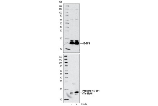


Phospho-4E-BP1 (Thr37/46) (236B4) Rabbit mAb


 下载产品说明书 下载SDS
 用小程序,查商品更便捷


 收藏
收藏
 对比
对比 咨询
咨询
Product Usage Information
| Application | Dilution |
|---|---|
| Western Blotting | 1:1000 |
| Immunohistochemistry (Paraffin) | 1:800 - 1:3200 |
| Immunofluorescence (Immunocytochemistry) | 1:200 - 1:800 |
| Flow Cytometry (Fixed/Permeabilized) | 1:50 - 1:200 |
Specificity/Sensitivity
物种反应性:
人, 小鼠, 大鼠, 猴, 黑腹果蝇
参考图片
Immunohistochemical analysis of paraffin-embedded human lymphoma using Phospho-4E-BP1 (Thr37/46) (236B4) Rabbit mAb.
使用Phospho-4E-BP1 (Thr37/46) (236B4) Rabbit mAb兔单抗,免疫组化分析人类淋巴瘤组织石蜡切片。
Immunohistochemical analysis using Phospho-4E-BP1 (Thr37/46) (236B4) Rabbit mAb on SignalSlide (TM) Phospho-Akt (Ser473) IHC Controls #8101 (paraffin-embedded LNCaP cells untreated (left) or LY294002-treated (right)).
使用Phospho-4E-BP1 (Thr37/46) (236B4) Rabbit mAb兔单抗,免疫组化分析SignalSlide (TM) Phospho-Akt (Ser473) IHC Controls #8101 (未处理(左图) 和LY294002处理 (右图)的石蜡包埋的LNCaP细胞。
Flow cytometric analysis of Jurkat cells, untreated (green) or LY294002, Wortmannin and U0126-treated (blue), using Phospho-4E-BP1 (Thr36/46) (236B4) Rabbit mAb compared to a nonspecific negative control antibody (red).
与阴性非特异的对照抗体(红色)比较,使用Phospho-4E-BP1 (Thr36/46) (236B4) Rabbit mAb兔单抗,流式细胞术分析Jurkat细胞,细胞分为未处理组(绿色)或LY294002、Wortmannin和U0126处理组(蓝色)。
Immunohistochemical analysis of paraffin-embedded human colon carcinoma using Phospho-4E-BP1 (Thr37/46) (236B4) Rabbit mAb.
使用Phospho-4E-BP1 (Thr37/46) (236B4) Rabbit mAb兔单抗,免疫组化分析人类结肠癌组织石蜡切片。
Immunohistochemical analysis of paraffin-embedded human colon carcinoma using Phospho-4E-BP1 (Thr37/46) (236B4) Rabbit mAb in the presence of control peptide (left) or Phospho-4E-BP1 (Thr37/46) Blocking Peptide #1052 (right).
使用Phospho-4E-BP1 (Thr37/46) (236B4) Rabbit mAb兔单抗,免疫组化分析人类结肠癌组织石蜡切片,其分别孵育对照多肽(左图)和Phospho-4E-BP1 (Thr37/46) Blocking Peptide #1052 (右图)。
Confocal immunofluorescent analysis of HeLa cells treated with LY294002 (left) or 20% serum (right) and labeled with Phospho-4E-BP1 (Thr37/46) (236B4) Rabbit mAb (green). Actin filaments have been labeled with Alexa Fluor® 555 phalloidin (red).
使用Phospho-4E-BP1 (Thr37/46) (236B4) Rabbit mAb兔单抗(绿色),共聚焦免疫荧光观察Miwi蛋白在HeLa细胞中定位,细胞分别使用LY294002处理(左图)或20%血清处理(右图)。Alexa Fluor® 555 phalloidin标记微丝蛋白(红色)。
Western blot analysis of extracts from 293T cells using 4E-BP1 Antibody #9452 (upper) and Phospho-4E-BP1 (Thr37/46) Antibody #2855 (lower). The cells were starved for 24 hours in serum-free medium and underwent a 1 hour amino acid deprivation. Amino acids were replenished for 1 hour. Cells were then either untreated (-) or treated with 100 nM insulin (+) for 30 minutes.
使用4E-BP1 Antibody #9452 (上图)和Phospho-4E-BP1 (Thr37/46) Antibody #2855 (下图),免疫印迹(Western Blot)分析293 T细胞系裂解物。细胞在无血清饥饿培养24小时,接着氨基酸缺乏培养1小时,然后再加入氨基酸培养1小时。最后,细胞加入100 nM胰岛素 (+)或不加(-)处理30分钟。