

 全部商品分类


1/2

品牌: BD Pharmingen
 下载产品说明书 下载SDS
 用小程序,查商品更便捷


 收藏
收藏
 对比
对比 咨询
咨询反应种属:
Rhesus, Cynomolgus, Baboon (QC Testing), Human (Tested in Development)
来源宿主:
Mouse IgG1, κ
产品介绍
产品信息
荧光素标记
抗原名称
CD8

宿主
Mouse IgG1, κ
免疫原
Human CD8a
简单描述
The RPA-T8 monoclonal antibody specifically binds to CD8 alpha (CD8α). CD8α is a type I transmembrane glycoprotein and a member of the immunoglobulin superfamily. CD8α is expressed by the majority of thymocytes, by subpopulations of αβ T cells and γδ T cells and by some NK cells. Cell surface CD8α is expressed either as a disulfide-linked homodimer (CD8αα) or as a heterodimer (CD8αβ) when disulfide-bonded to a CD8 beta chain (CD8β). CD8-positive αβ T cells coexpress both CD8αα homodimers and CD8αβ heterodimers whereas some γδ T cells and NK cells express CD8αα homodimers. CD8 plays important roles in T cell activation and selection. The extracellular IgSF domain of CD8α binds to a non-polymorphic determinant on HLA class I molecules (α3 domain) and enables CD8 to function as a co-receptor with MHC class I-restricted TCR during T cell recognition of antigen. The cytoplasmic domain of CD8α associates with Lck, a Src family protein tyrosine kinase that is involved in intracellular signaling. The RPA-T8 and HIT8a monoclonal antibodies are not cross-blocking. This clone has been reported to react with a subset of peripheral blood lymphocytes, but not monocytes nor granuloyctes, of baboon and both rhesus and cynomolgus macaque monkey. In general, a higher frequency of CD8+ and CD4+CD8+ lymphocytes are observed in non-human primates compared to normal human donors.
商品描述
RPA-T8
The RPA-T8 monoclonal antibody specifically binds to CD8 alpha (CD8α). CD8α is a type I transmembrane glycoprotein and a member of the immunoglobulin superfamily. CD8α is expressed by the majority of thymocytes, by subpopulations of αβ T cells and γδ T cells and by some NK cells. Cell surface CD8α is expressed either as a disulfide-linked homodimer (CD8αα) or as a heterodimer (CD8αβ) when disulfide-bonded to a CD8 beta chain (CD8β). CD8-positive αβ T cells coexpress both CD8αα homodimers and CD8αβ heterodimers whereas some γδ T cells and NK cells express CD8αα homodimers. CD8 plays important roles in T cell activation and selection. The extracellular IgSF domain of CD8α binds to a non-polymorphic determinant on HLA class I molecules (α3 domain) and enables CD8 to function as a co-receptor with MHC class I-restricted TCR during T cell recognition of antigen. The cytoplasmic domain of CD8α associates with Lck, a Src family protein tyrosine kinase that is involved in intracellular signaling. The RPA-T8 and HIT8a monoclonal antibodies are not cross-blocking. This clone has been reported to react with a subset of peripheral blood lymphocytes, but not monocytes nor granuloyctes, of baboon and both rhesus and cynomolgus macaque monkey. In general, a higher frequency of CD8+ and CD4+CD8+ lymphocytes are observed in non-human primates compared to normal human donors.
同种型
Mouse IgG1, κ
克隆号
克隆 RPA-T8 (RUO)
产品详情
APC
Allophycocyanin (APC), is part of the BD family of phycobiliprotein dyes. This fluorochrome is a multimeric fluorescent phycobiliprotein with excitation maximum (Ex Max) of 651 nm and an emission maximum (Em Max) at 660 nm. APC is designed to be excited by the Red (627-640 nm) laser and detected using an optical filter centered near 660 nm (e.g., a 660/20 nm bandpass filter). Please ensure that your instrument’s configurations (lasers and optical filters) are appropriate for this dye.

APC
Red 627-640 nm
651 nm
660 nm
应用
实验应用
Flow cytometry (Routinely Tested)
推荐用量
5 µl
反应种属
Rhesus, Cynomolgus, Baboon (QC Testing), Human (Tested in Development)
目标/特异性
CD8
背景
别名
CD8α; CD8A; CD8 alpha; Leu2; MAL; T8; p32
制备和贮存
存储溶液
Aqueous buffered solution containing BSA and ≤0.09% sodium azide.
保存方式
Aqueous buffered solution containing BSA and ≤0.09% sodium azide.
文献
文献
研发参考(5)
1. Knapp W. W. Knapp .. et al., ed. Leucocyte typing IV : white cell differentiation antigens. Oxford New York: Oxford University Press; 1989:1-1182.
2. Reimann KA, Waite BC, Lee-Parritz DE, et al. Use of human leukocyte-specific monoclonal antibodies for clinically immunophenotyping lymphocytes of rhesus monkeys. Cytometry. 1994; 17(1):102-108. (Biology).
3. Schlossman SF. Stuart F. Schlossman .. et al., ed. Leucocyte typing V : white cell differentiation antigens : proceedings of the fifth international workshop and conference held in Boston, USA, 3-7 November, 1993. Oxford: Oxford University Press; 1995.
4. Schlossman SF. Stuart F. Schlossman .. et al., ed. Leucocyte typing V : white cell differentiation antigens : proceedings of the fifth international workshop and conference held in Boston, USA, 3-7 November, 1993. Oxford: Oxford University Press; 1995.
5. Sopper S, Stahl-Hennig C, Demuth M, Johnston IC, Dorries R, ter Meulen V. Lymphocyte subsets and expression of differentiation markers in blood and lymphoid organs of rhesus monkeys. Cytometry. 1997; 29(4):351-362. (Biology).
数据库链接
Entrez-Gene ID
925
参考图片
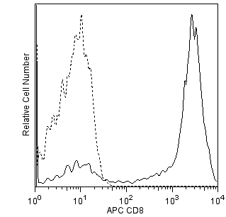
Flow cytometric analysis of CD8 expression on Rhesus macaque peripheral blood lymphocytes. Rhesus macaque whole blood was stained with APC Mouse anti-Human CD8 antibody (Cat. No. 561421/561952/561953; solid line histogram) or with an APC Mouse IgG1, κ Isotype Control (Cat. No. 554681; dashed line histogram). The erythrocytes were lysed with BD PharmLyse™ Lysing Buffer (Cat. No. 555899). The fluorescence histograms were derived from events with the forward and side light-scatter characteristics of viable lymphocytes. Flow cytometry was performed using a BD™ LSR II Flow Cytometer System.
声明 :本官网所有报价均为常温或者蓝冰运输价格,如有产品需要干冰运输,需另外加收干冰运输费。