


 全部商品分类
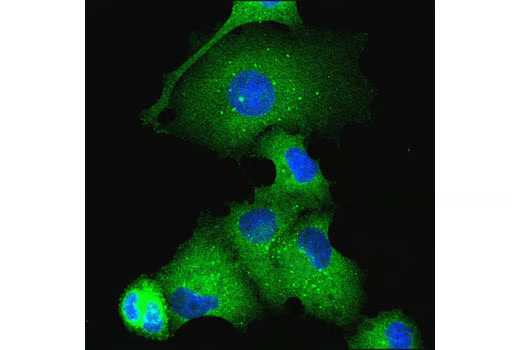


Atg16L1 (D6D5) Rabbit mAb

 下载产品说明书 下载COA 下载SDS
 用小程序,查商品更便捷


 收藏
收藏
 对比
对比 咨询
咨询
Monoclonal antibody is produced by immunizing animals with a synthetic peptide corresponding to residues surrounding Val51 of human Atg16L1 protein.
Product Usage Information
| Application | Dilution |
|---|---|
| Western Blotting | 1:1000 |
| Immunoprecipitation | 1:100 |
| Immunofluorescence (Immunocytochemistry) | 1:50 - 1:200 |
Specificity/Sensitivity
Species Reactivity:
Human, Mouse, Rat
Supplied in 10 mM sodium HEPES (pH 7.5), 150 mM NaCl, 100 µg/ml BSA, 50% glycerol and less than 0.02% sodium azide. Store at –20°C. Do not aliquot the antibody.
参考图片
Confocal immunofluorescent analysis of EBSS-starved PANC-1 cells using Atg16L1 (D6D5) Rabbit mAb (green). Blue pseudocolor = DRAQ5® #4084 (fluorescent DNA dye).
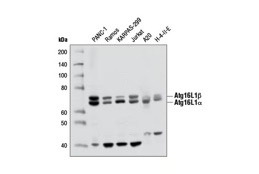
Western blot analysis of extracts from various cell lines using Atg16L1 (D6D5) Rabbit mAb.
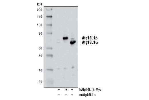
Western blot analysis of extracts from 293T cells, mock transfected (-) or transfected with either a myc-tagged human Atg16L1β construct (hAtg16L1β-Myc; +) or a mouse Atg16L1α construct (mAtg16L1α; +), using Atg16L1 (D6D5) Rabbit mAb. The myc-tagged human Atg16L1β construct was kindly provided by Dr. Qing Zhong, University of California Berkeley.
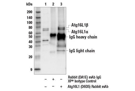
Immunoprecipitation of Atg16L1 from Jurkat cell extracts. Lane 1 is 10% input, lane 2 is precipitated with Rabbit (DA1E) mAb IgG XP® Isotype Control #3900, and lane 3 is Atg16L1 (D6D5) Rabbit mAb, #8089. Western blot was performed using Atg16L1 (D6D5) Rabbit mAb.