


 全部商品分类


AIF (D39D2) XP ® Rabbit mAb

 下载产品说明书 下载COA 下载SDS
 用小程序,查商品更便捷


 收藏
收藏
 对比
对比 咨询
咨询
Monoclonal antibody was produced by immunizing animals with a synthetic peptide corresponding to residues surrounding Ala520 of human AIF protein.
Product Usage Information
| Application | Dilution |
|---|---|
| Western Blotting | 1:1000 |
| Immunoprecipitation | 1:100 |
| Immunohistochemistry (Paraffin) | 1:50 - 1:200 |
| Immunofluorescence (Frozen) | 1:200 - 1:400 |
| Immunofluorescence (Immunocytochemistry) | 1:200 - 1:400 |
Specificity/Sensitivity
Species Reactivity:
Human, Mouse, Rat, Monkey
Supplied in 10 mM sodium HEPES (pH 7.5), 150 mM NaCl, 100 µg/ml BSA, 50% glycerol and less than 0.02% sodium azide. Store at –20°C. Do not aliquot the antibody.
For a carrier free (BSA and azide free) version of this product see product #62147.
参考图片
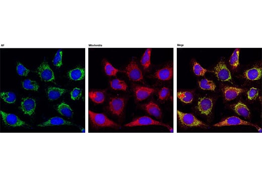
Confocal immunofluorescent analysis of HeLa cells using AIF (D39D2) XP ™ Rabbit mAb (green), showing colocalization with mitochondria that have been labeled with MitoTracker® Red CMXRos (red). Blue pseudocolor = DRAQ5® #4084 (fluorescent DNA dye).
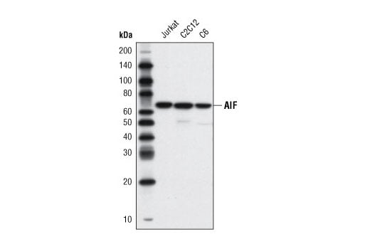
Western blot analysis of extracts from Jurkat, C2C12, and C6 cells using AIF (D39D2) XP® Rabbit mAb.
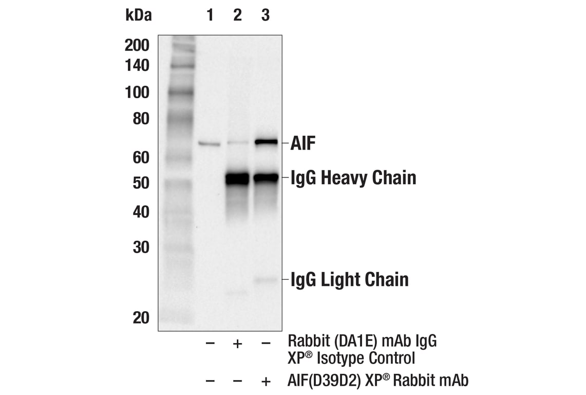
Immunoprecipitation of AIF protein from Jurkat cell extracts. Lane 1 is 10% input, lane 2 is Rabbit (DA1E) mAb IgG XP® Isotype Control #3900, and lane 3 is AIF (D39D2) XP® Rabbit mAb. Western blot analysis was performed using AIF (D39D2) XP® Rabbit mAb. Anti-rabbit IgG, HRP-linked Antibody #7074 was used as a secondary antibody.
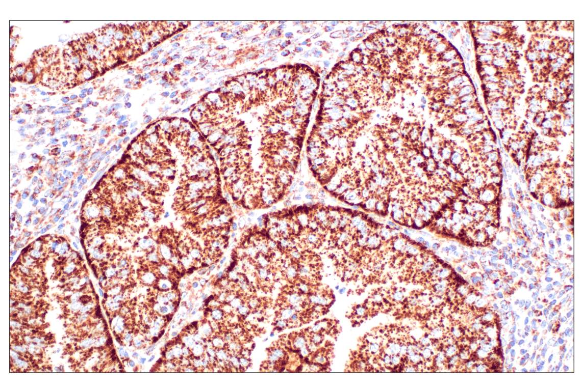
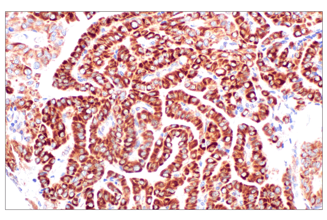
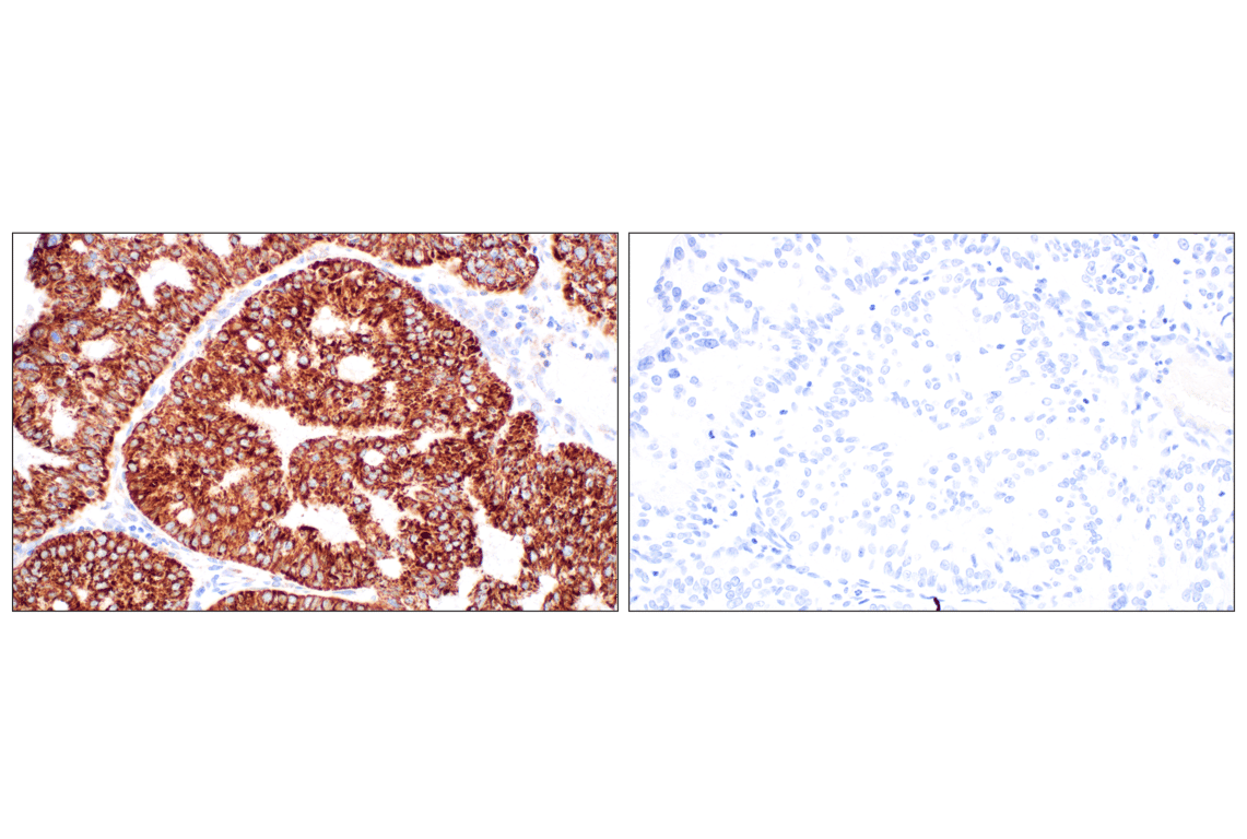
Immunohistochemical analysis of paraffin-embedded human endometrioid adenocarcinoma using AIF (D39D2) XP® Rabbit mAb.
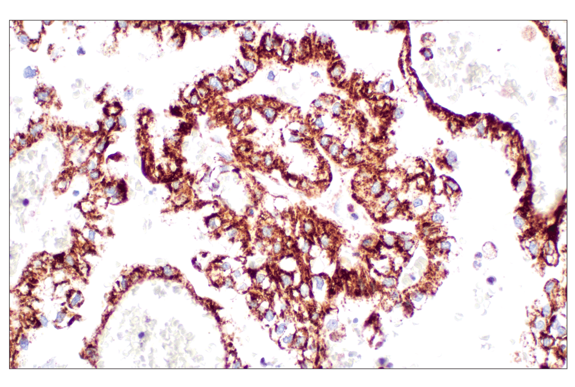
Immunohistochemical analysis of paraffin-embedded human papillary thyroid carcinoma using AIF (D39D2) XP® Rabbit mAb.
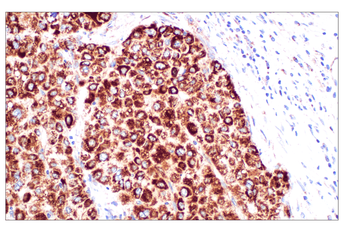
Immunohistochemical analysis of paraffin-embedded human hepatocellular carcinoma using AIF (D39D2) XP® Rabbit mAb.
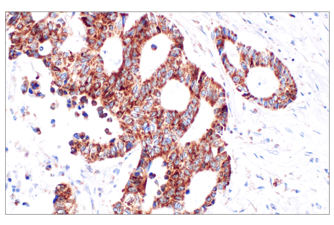
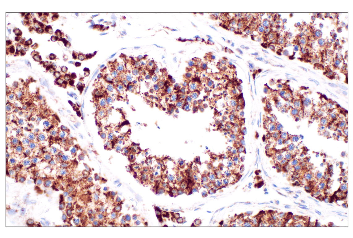
Immunohistochemical analysis of paraffin-embedded human renal cell carcinoma using AIF (D39D2) XP® Rabbit mAb.
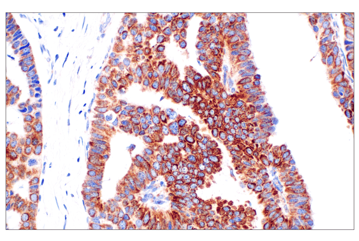
Immunohistochemical analysis of paraffin-embedded human colon adenocarcinoma using AIF (D39D2) XP® Rabbit mAb.
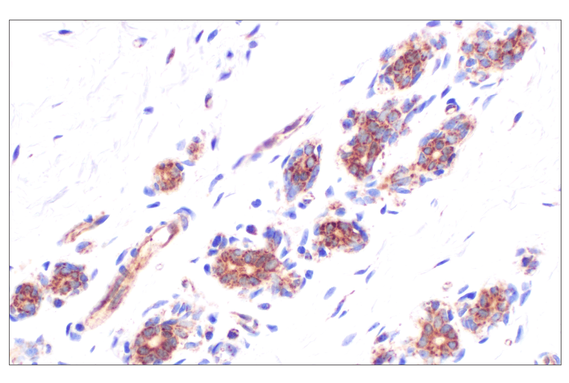
Immunohistochemical analysis of paraffin-embedded human papillary carcinoma of the breast using AIF (D39D2) XP® Rabbit mAb.
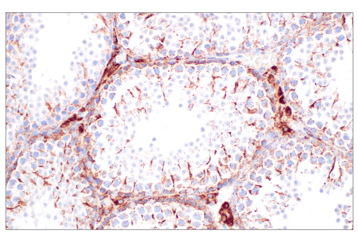
Immunohistochemical analysis of paraffin-embedded normal human testis using AIF (D39D2) XP® Rabbit mAb.
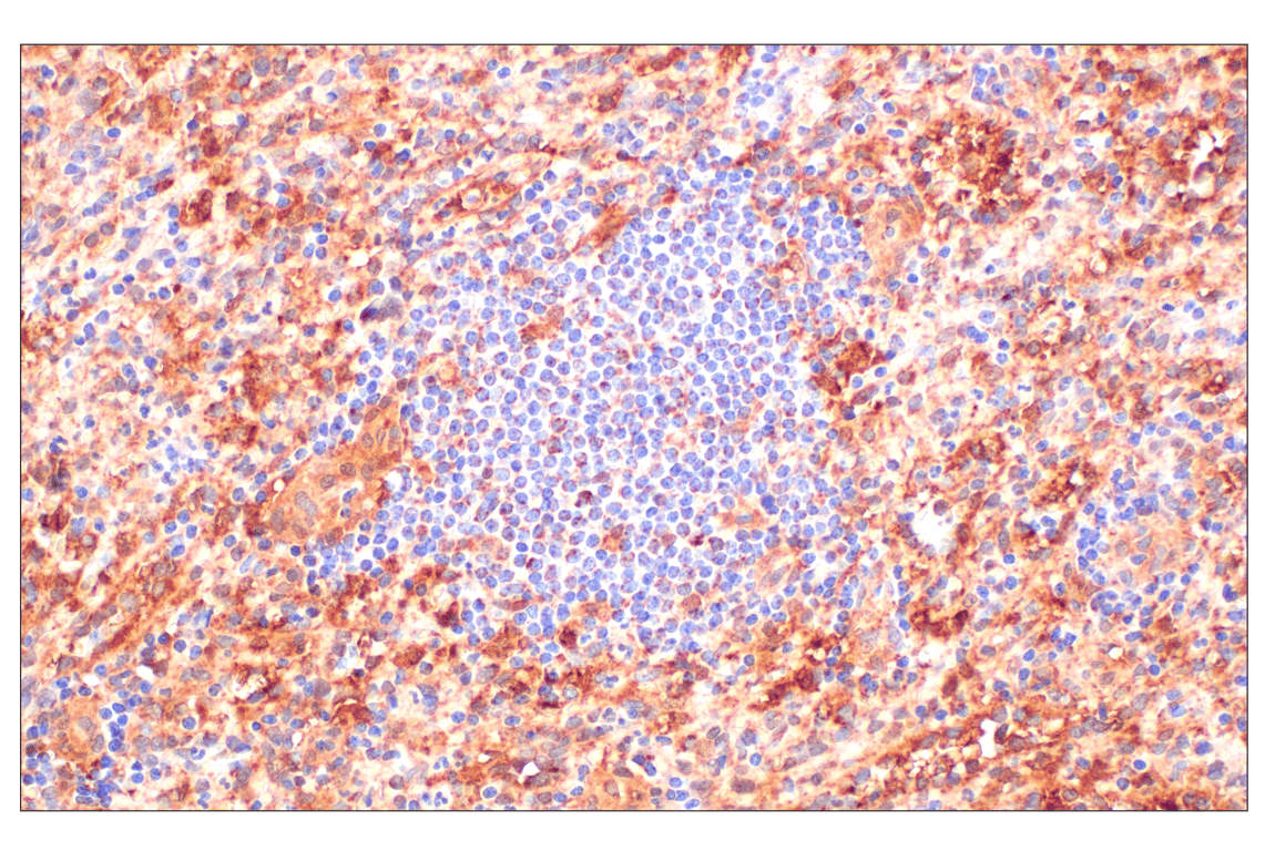
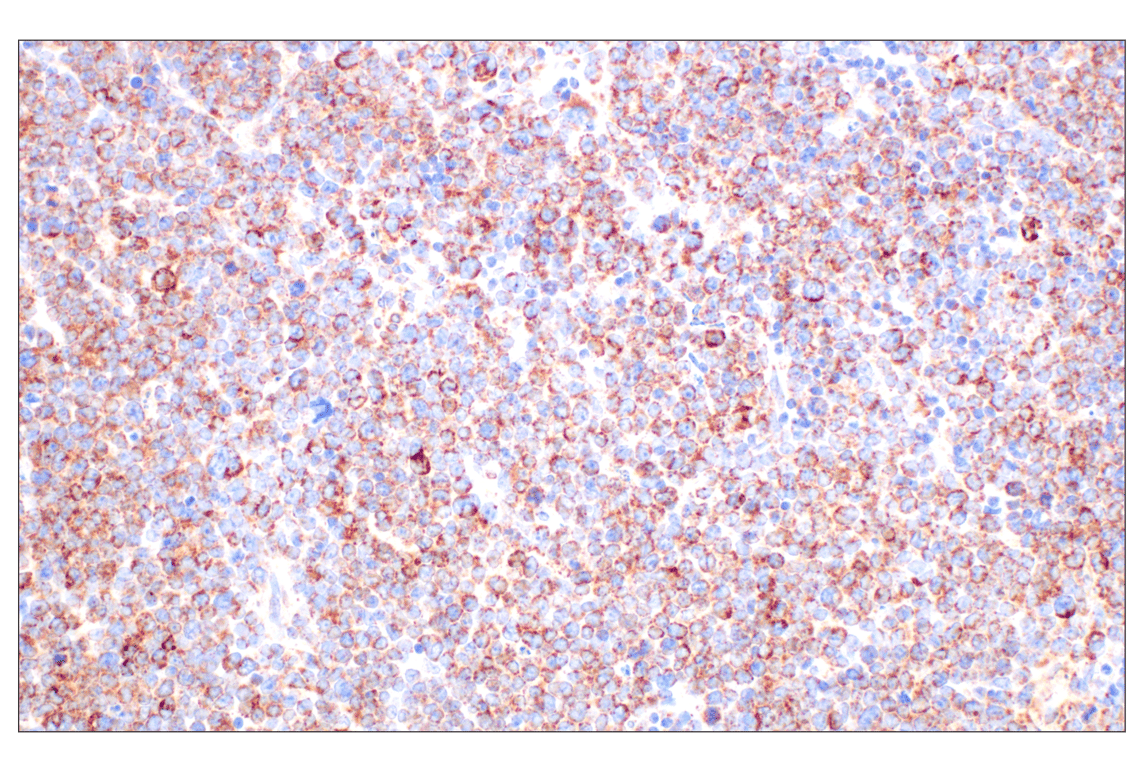
Immunohistochemical analysis of paraffin-embedded normal human spleen using AIF (D39D2) XP® Rabbit mAb.
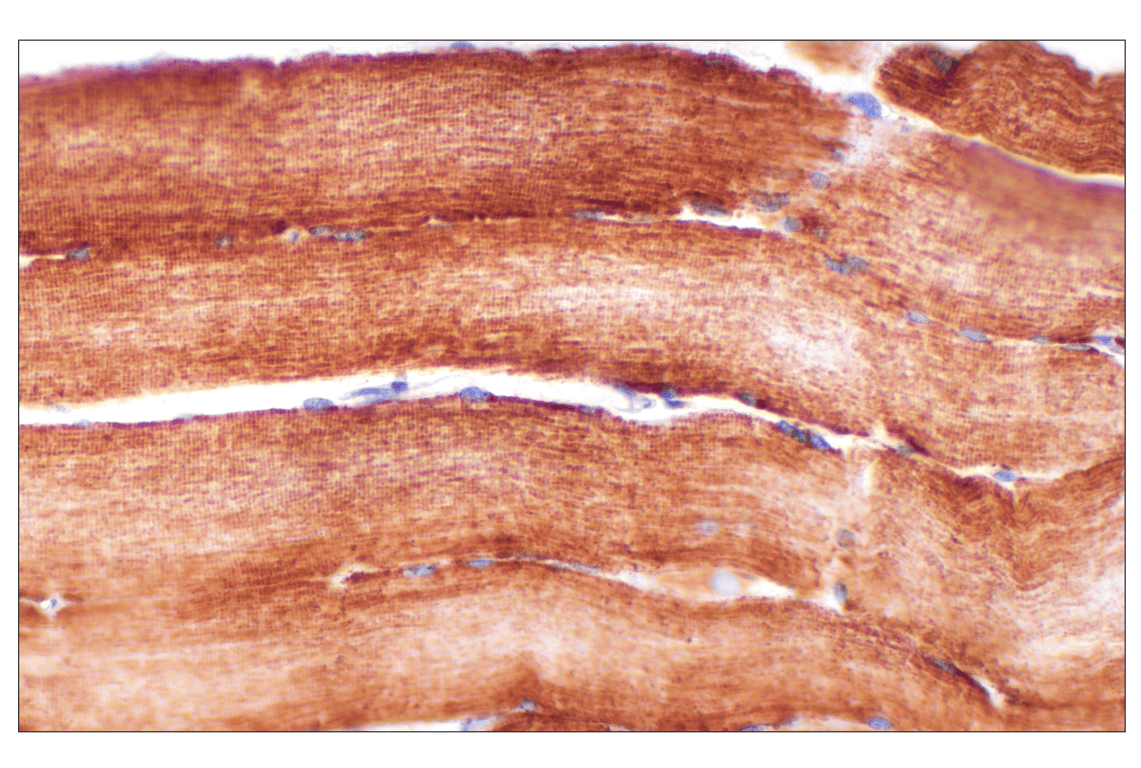
Immunohistochemical analysis of paraffin-embedded normal human skeletal muscle using AIF (D39D2) XP® Rabbit mAb.
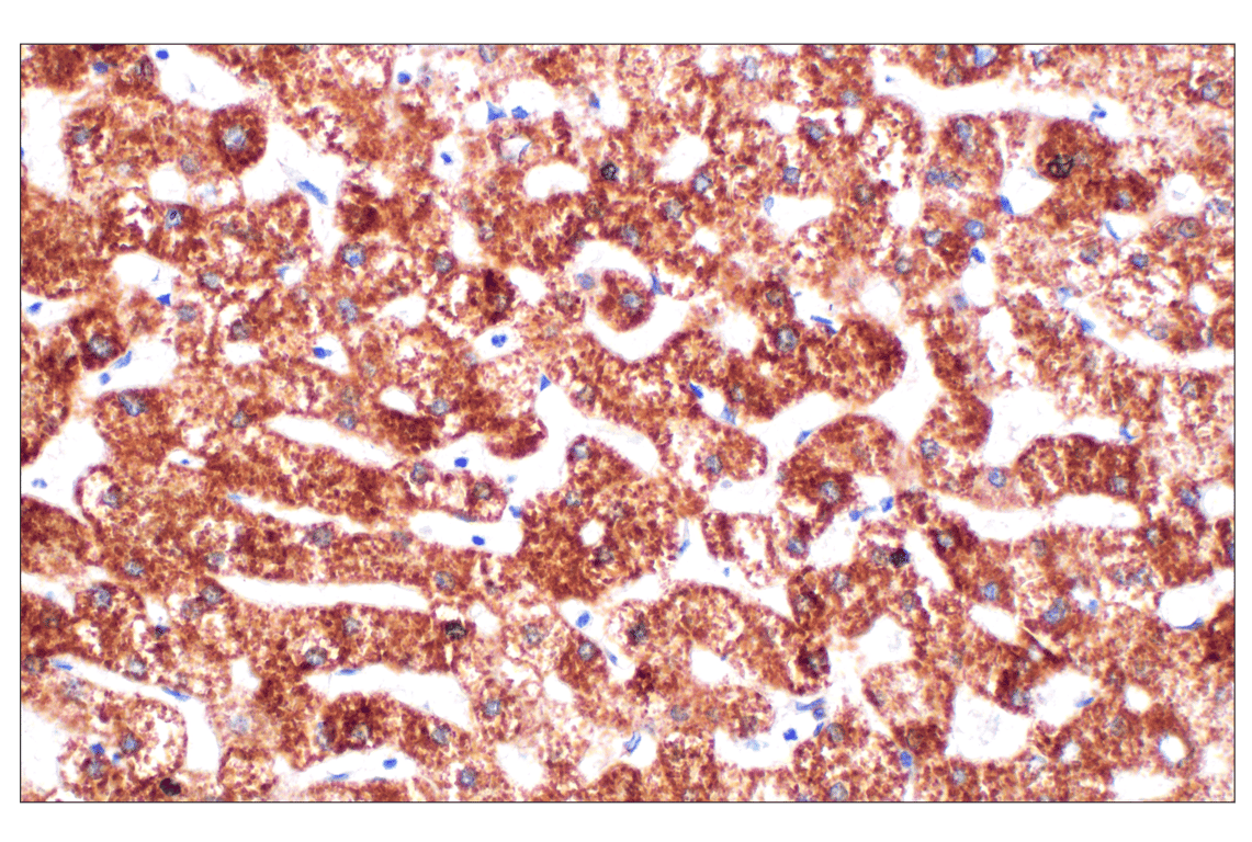
Immunohistochemical analysis of paraffin-embedded normal human liver using AIF (D39D2) XP® Rabbit mAb.
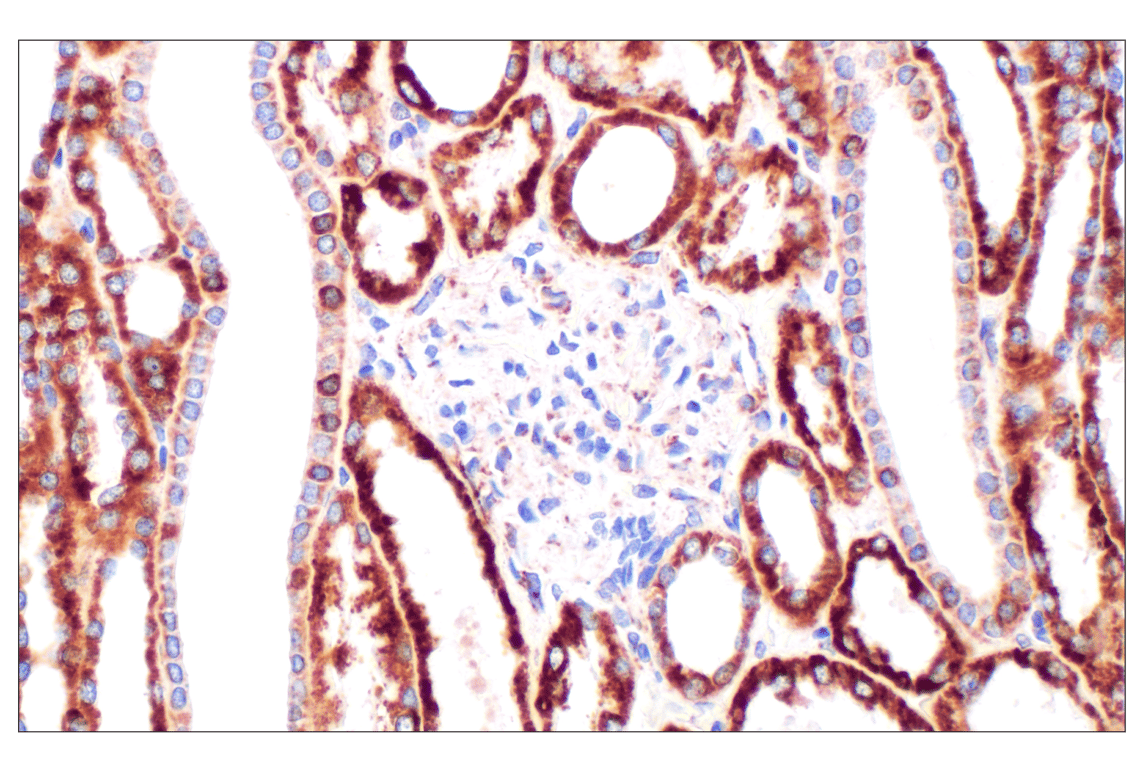
Immunohistochemical analysis of paraffin-embedded normal human kidney using AIF (D39D2) XP® Rabbit mAb.
Immunohistochemical analysis of paraffin-embedded normal human breast using AIF (D39D2) XP® Rabbit mAb.
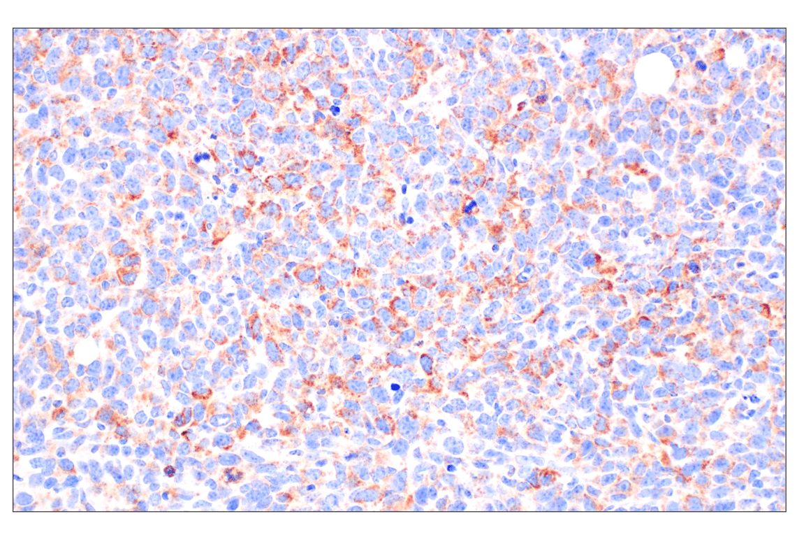
Immunohistochemical analysis of paraffin-embedded A20 syngeneic tumor using AIF (D39D2) XP® Rabbit mAb.
Immunohistochemical analysis of paraffin-embedded LL/2 syngeneic tumor using AIF (D39D2) XP® Rabbit mAb.
Immunohistochemical analysis of paraffin-embedded mouse testis using AIF (D39D2) XP® Rabbit mAb.
Immunohistochemical analysis of paraffin-embedded human endometrioid adenocarcinoma using AIF (D39D2) XP® Rabbit mAb (left) compared to concentration-matched Rabbit (DA1E) mAb IgG XP® Isotype Control #3900 (right).
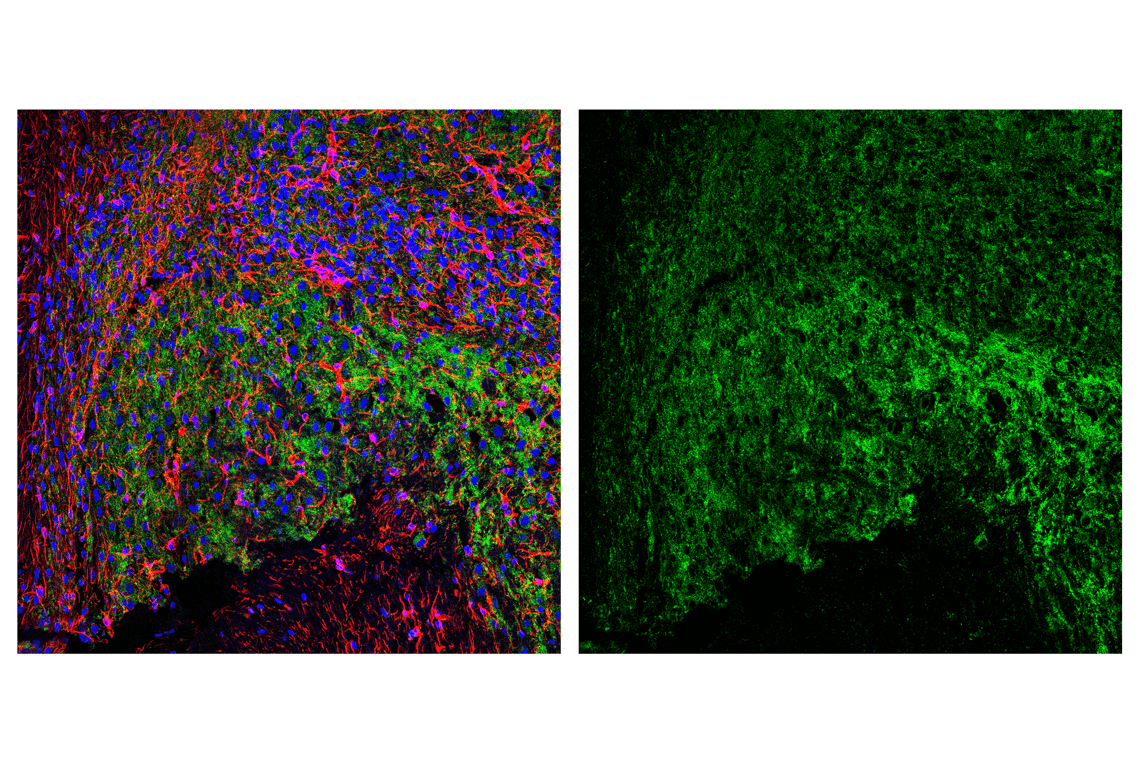
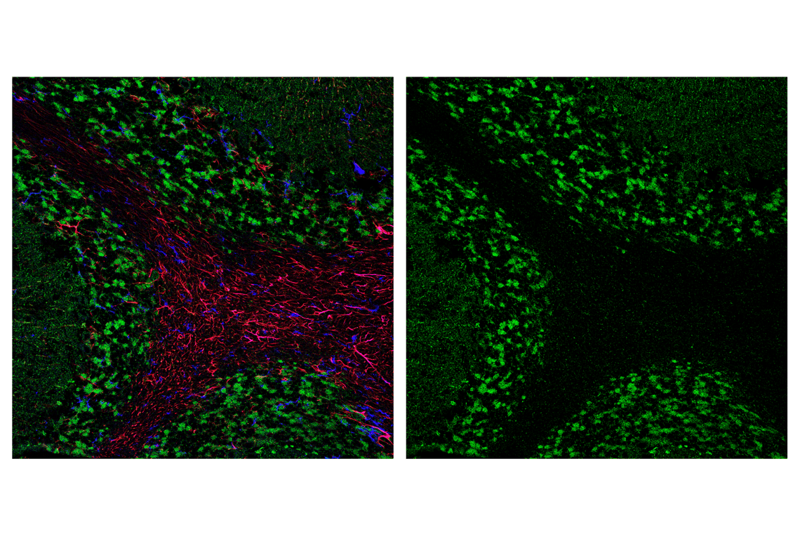
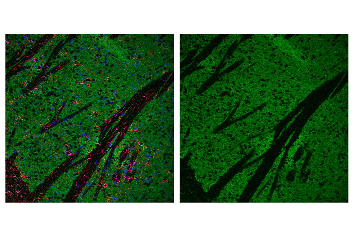
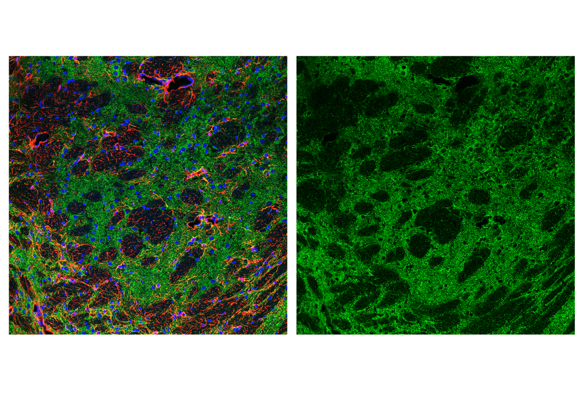
Confocal immunofluorescent analysis of mouse brainstem using AIF (D39D2) XP® Rabbit mAb #5318 (green) and GFAP (GA5) Mouse mAb #3670 (red). Sections were mounted in ProLong® Gold Antifade Reagent with DAPI #8961 (blue).
Confocal immunofluorescent analysis of mouse cerebellum using AIF (D39D2) XP® Rabbit mAb #5318 (green), GFAP (GA5) Mouse mAb #3670 (red), and F4/80 (BM8.1) Rat mAb #71299 (blue).
Confocal immunofluorescent analysis of mouse striatum using AIF (D39D2) XP® Rabbit mAb #5318 (green), GFAP (GA5) Mouse mAb #3670 (red), and F4/80 (BM8.1) Rat mAb #71299 (blue).
Confocal immunofluorescent analysis of mouse thalamus using AIF (D39D2) XP® Rabbit mAb #5318 (green), GFAP (GA5) Mouse mAb #3670 (red). Sections were mounted in ProLong® Gold Antifade Reagent with DAPI #8961 (blue).
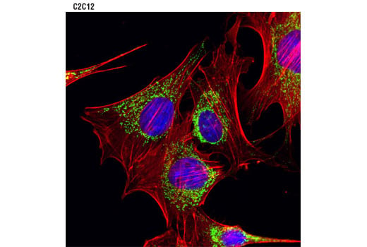
Confocal immunofluorescent analysis of C2C12 cells using AIF (D39D2) XP® Rabbit mAb #5318 (green). Actin filaments were labeled with DY-554 phalloidin (red). Blue pseudocolor = DRAQ5® #4084 (fluorescent DNA dye).