


 全部商品分类
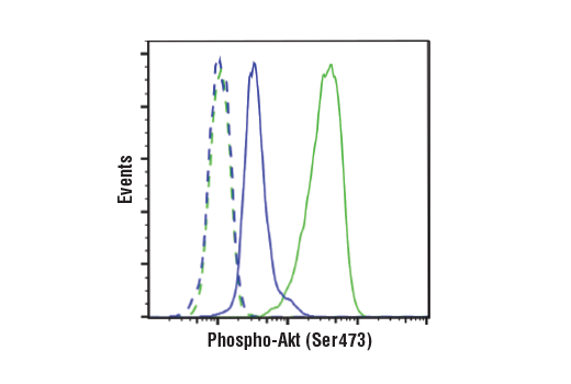


Phospho-Akt (Ser473) (D9E) XP® Rabbit mAb


 下载产品说明书 下载COA 下载SDS
 用小程序,查商品更便捷


 收藏
收藏
 对比
对比 咨询
咨询
Monoclonal antibody is produced by immunizing animals with a synthetic phosphopeptide corresponding to residues around Ser473 of human Akt.
Product Usage Information
| Application | Dilution |
|---|---|
| Western Blotting | 1:2000 |
| Fluorescent Western | 1:2000 |
| Simple Western™ | 1:10 - 1:50 |
| Immunoprecipitation | 1:50 |
| Immunohistochemistry (Paraffin) | 1:50 - 1:200 |
| Immunofluorescence (Immunocytochemistry) | 1:400 - 1:800 |
| Flow Cytometry (Fixed/Permeabilized) | 1:100 - 1:400 |
Specificity/Sensitivity
Species Reactivity:
Human, Mouse, Rat, Hamster, Monkey, D. melanogaster, Zebrafish, Bovine
Supplied in 10 mM sodium HEPES (pH 7.5), 150 mM NaCl, 100 µg/ml BSA, 50% glycerol and less than 0.02% sodium azide. Store at –20°C. Do not aliquot the antibody.
For a carrier-free (BSA and azide free) version of this product see product #31957.
参考图片
Flow cytometric analysis of Jurkat cells, untreated (green) or treated with LY294002 #9901, Wortmannin #9951, and U0126 #9903 (50 μM, 1 μM, and 10 μM, 2 hr; blue) using Phospho-Akt (Ser473) (D9E) XP® Rabbit mAb (solid lines) or concentration-matched Rabbit (DA1E) mAb IgG XP® Isotype Control #3900 (dashed lines). Anti-rabbit IgG (H+L), F(ab')2 Fragment (Alexa Fluor® 488 Conjugate) #4412 was used as a secondary antibody.
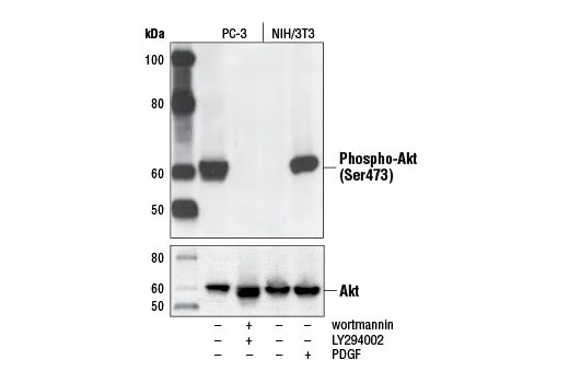
Western blot analysis of extracts from PC-3 cells, untreated or LY294002/wortmannin-treated, and NIH/3T3 cells, serum-starved or PDGF-treated, using Phospho-Akt (Ser473) (D9E) XP® Rabbit mAb (upper) or Akt (pan) (C67E7) Rabbit mAb #4691 (lower).
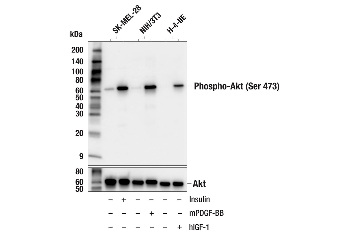
Western blot analysis of extracts from various cell lines, untreated (-) or treated (+) as indicated with human insulin (100 nM, 20 min), mouse PDGF-BB (100 ng/ml, 20 min), or human Insulin-like Growth Factor I (hIGF-I) #8917 (100 ng/ml; 5 min), using Phospho-Akt (Ser473) (D9E) XP® Rabbit mAb (upper) or Akt (pan) (C67E7) Rabbit mAb #4691 (lower).
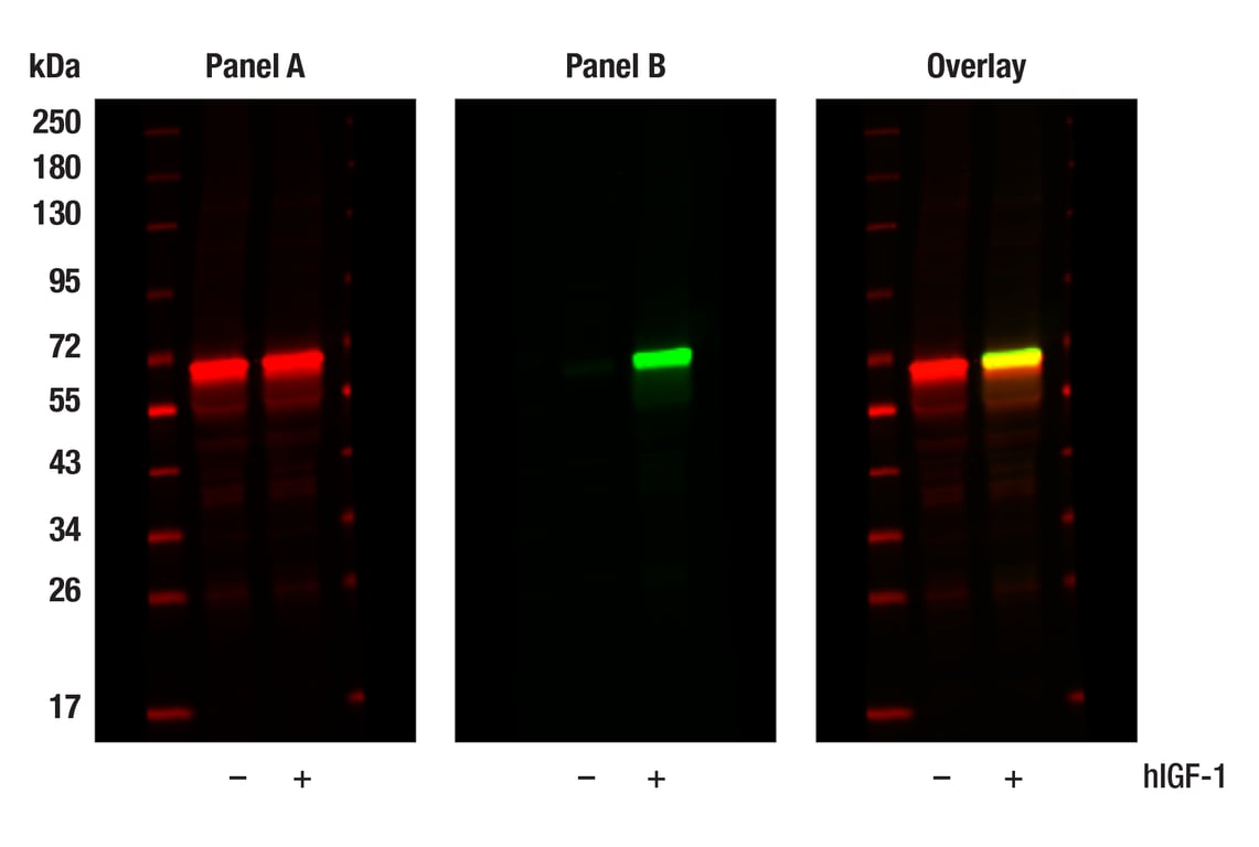
Western blot analysis of extracts from MCF-7 cells, untreated (-) or treated with hIGF-1 (100 ng/ml, 10 min; +), using Akt (pan) (E7J2C) Mouse mAb #58295 (Panel A) and Phospho-Akt (Ser473) (D9E) XP® Rabbit mAb #4060 (Panel B). Anti-mouse IgG (H+L) (DyLight 680 Conjugate) #5470 (red) and Anti-rabbit IgG (H+L) (DyLight 800 4X PEG Conjugate) #5151 (green) were used as secondary antibodies.
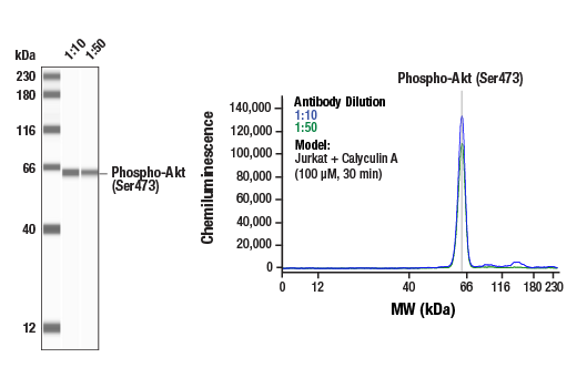
Simple Western™ analysis of lysates (0.1 mg/mL) from Jurkat cells treated with Calyculin A (100 uM, 30 min) using Phospho-Akt (Ser473) (D9E) XP® Rabbit mAb #4060. The virtual lane view (left) shows a single target band (as indicated) at 1:10 and 1:50 dilutions of primary antibody. The corresponding electropherogram view (right) plots chemiluminescence by molecular weight along the capillary at 1:10 (blue line) and 1:50 (green line) dilutions of primary antibody. This experiment was performed under reducing conditions on the Jess™ Simple Western instrument from ProteinSimple, a BioTechne brand, using the 12-230 kDa separation module.
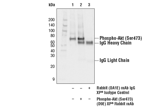
Immunoprecipitation of phospho-Akt (Ser473) from Jurkat extracts treated with Calyculin A #9902 (100nM, 30 min). Lane 1 is 10% input, lane 2 is Phospho-Akt (Ser473) (D9E) XP® Rabbit mAb, and lane 3 is Rabbit (DA1E) mAb IgG XP® Isotype Control #3900. Western blot analysis was performed with Phospho-Akt (Ser473) (D9E) XP® Rabbit mAb. Anti-rabbit IgG, HRP-linked Antibody #7074 was used as a secondary antibody.
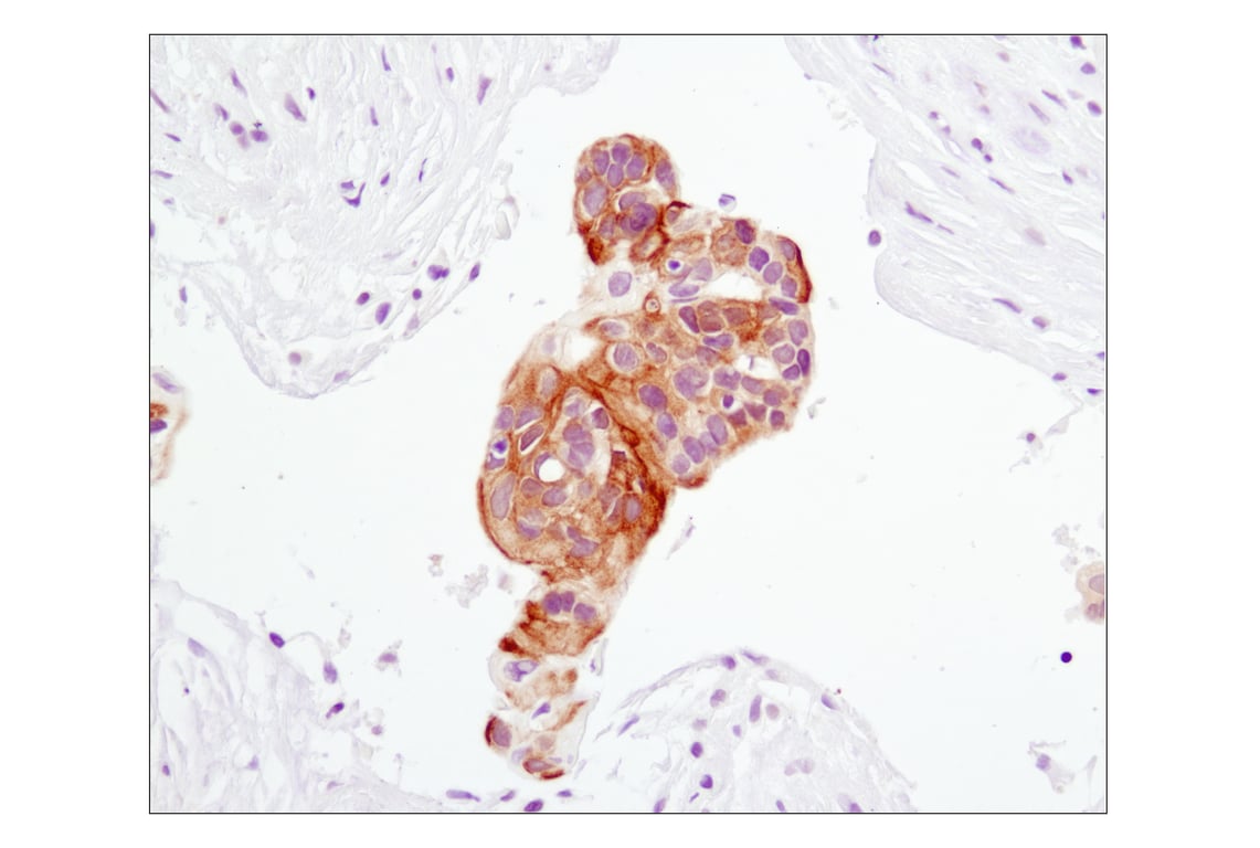
Immunohistochemical analysis of paraffin-embedded human lung carcinoma using Phospho-Akt (Ser473) (D9E) XP® Rabbit mAb.
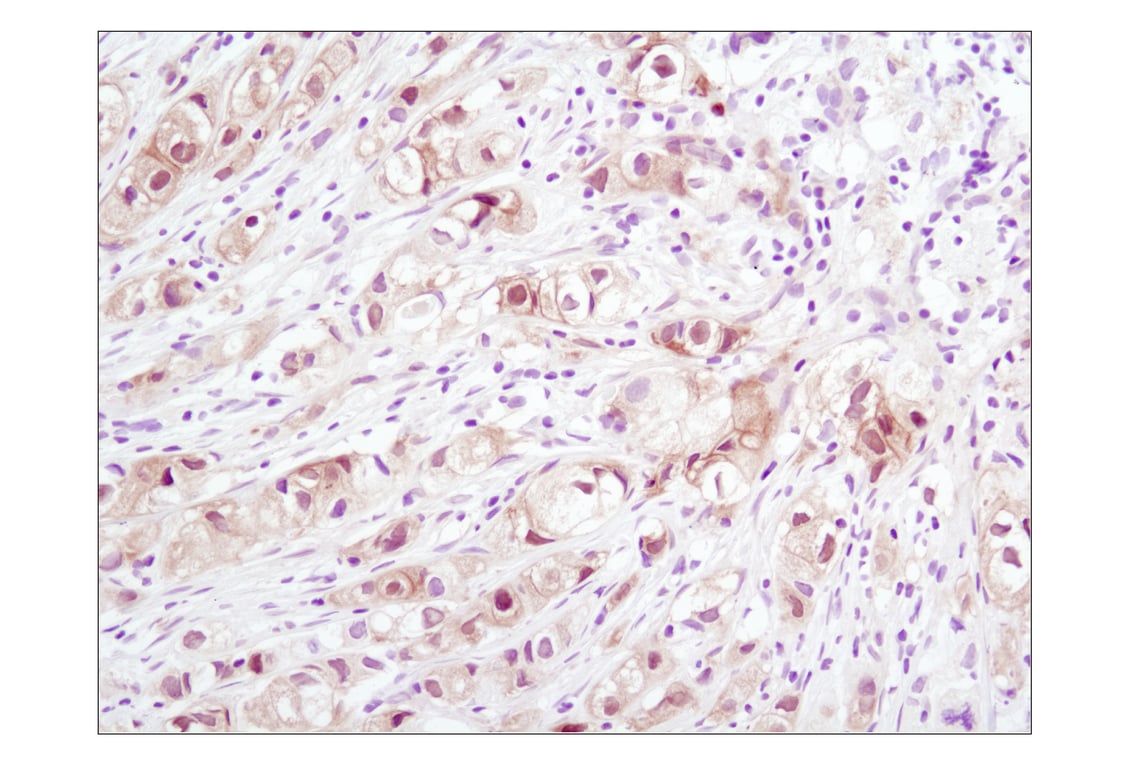
Immunohistochemical analysis of paraffin-embedded human breast carcinoma using Phospho-Akt (Ser473) (D9E) XP® Rabbit mAb.
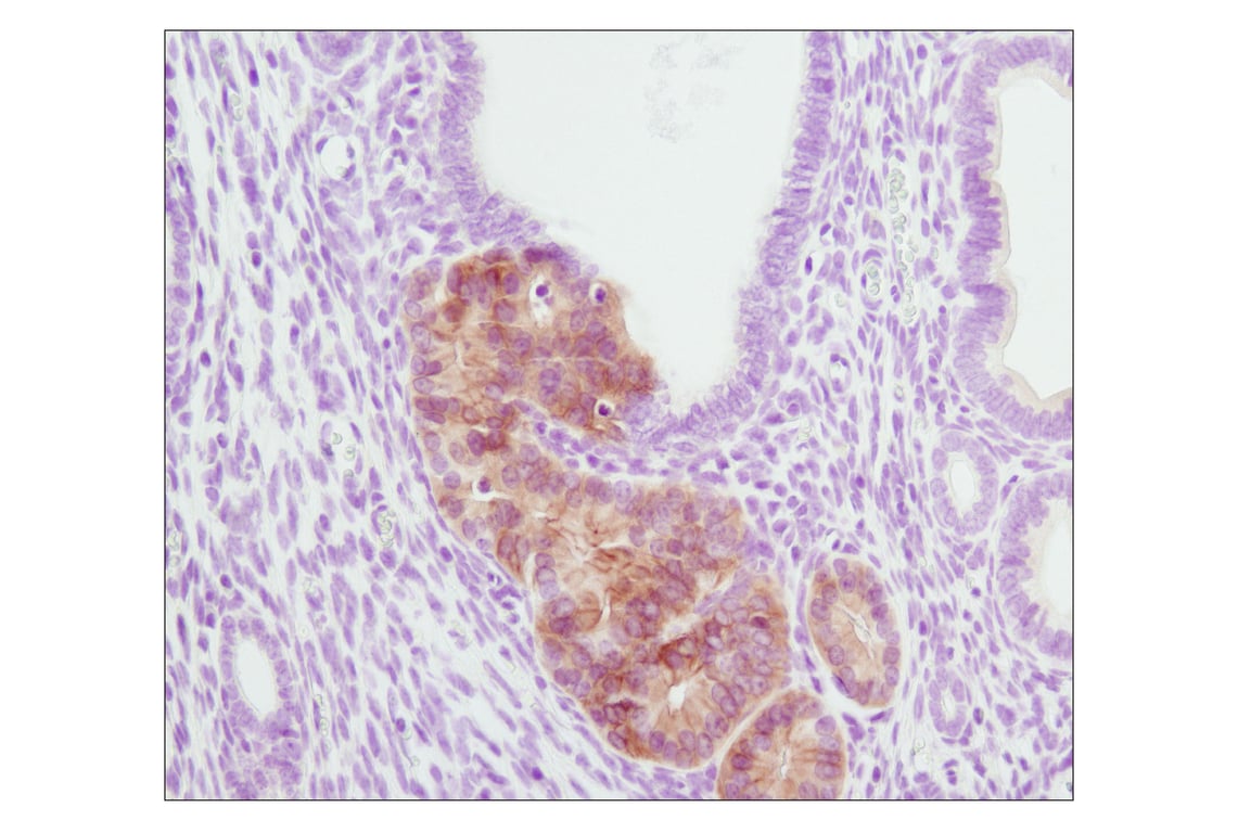
Immunohistochemical analysis of paraffin-embedded PTEN heterozygous mutant mouse endometrium using Phospho-Akt (Ser473) (D9E) XP® Rabbit mAb. (Tissue section courtesy of Dr. Sabina Signoretti, Brigham and Women's Hospital, Harvard Medical School, Boston, MA.)
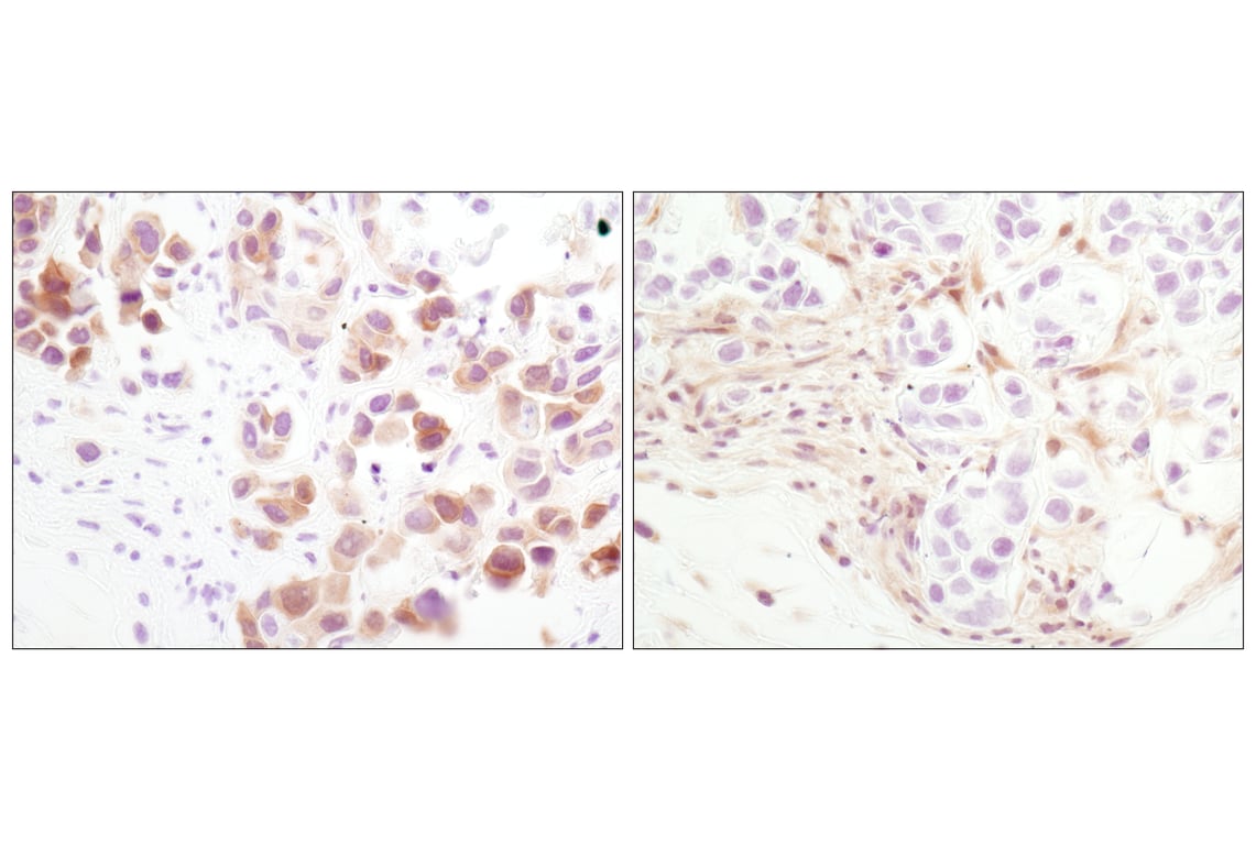
Immunohistochemical analysis of paraffin-embedded MDA-MB-468 xenograft using Phospho-Akt (Ser473) (D9E) XP® Rabbit mAb (left) or PTEN (138G6) Rabbit mAb #9559 (right). Note the presence of P-Akt staining in the PTEN deficient MDA-MB-468 cells.
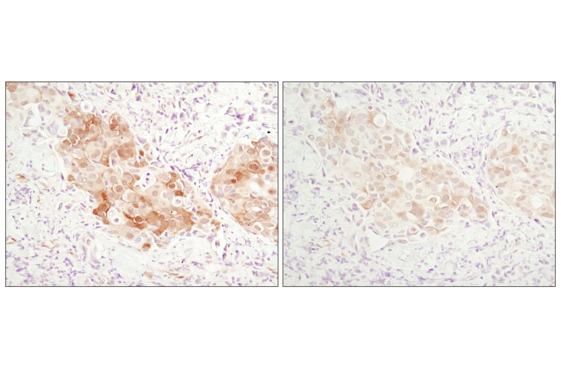
Immunohistochemical analysis of paraffin-embedded human breast carcinoma comparing SignalStain® Antibody Diluent #8112 (left) to TBST/5% normal goat serum (right) using Phospho-Akt (Ser473) (D9E) XP® Rabbit mAb #4060.
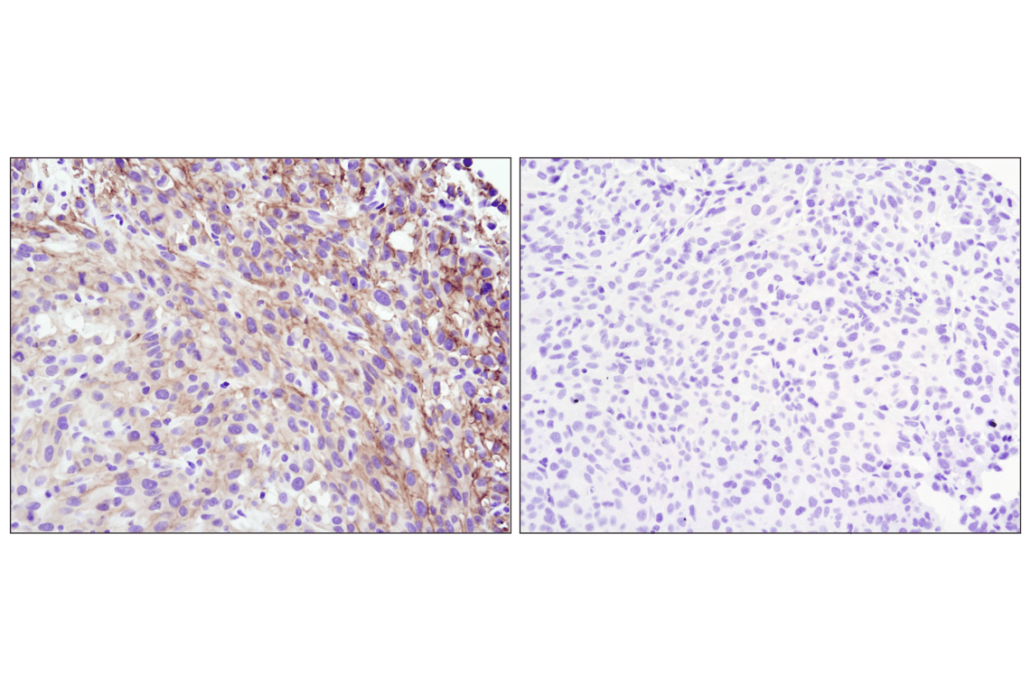
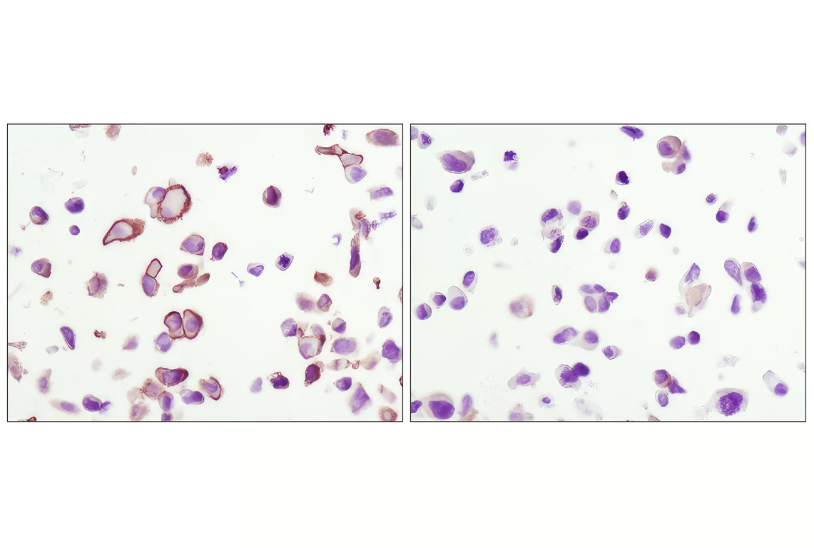
Immunohistochemical analysis of paraffin-embedded U-87MG xenograft, untreated (left) or lambda phosphatase-treated (right), using Phospho-Akt (Ser473) (D9E) XP® Rabbit mAb.
Immunohistochemical analysis using Phospho-Akt (Ser473) (D9E) XP® Rabbit mAb on SignalSlide® Phospho-Akt (Ser473) IHC Controls #8101 (paraffin-embedded LNCaP cells, untreated (left) or LY294002-treated (right)).
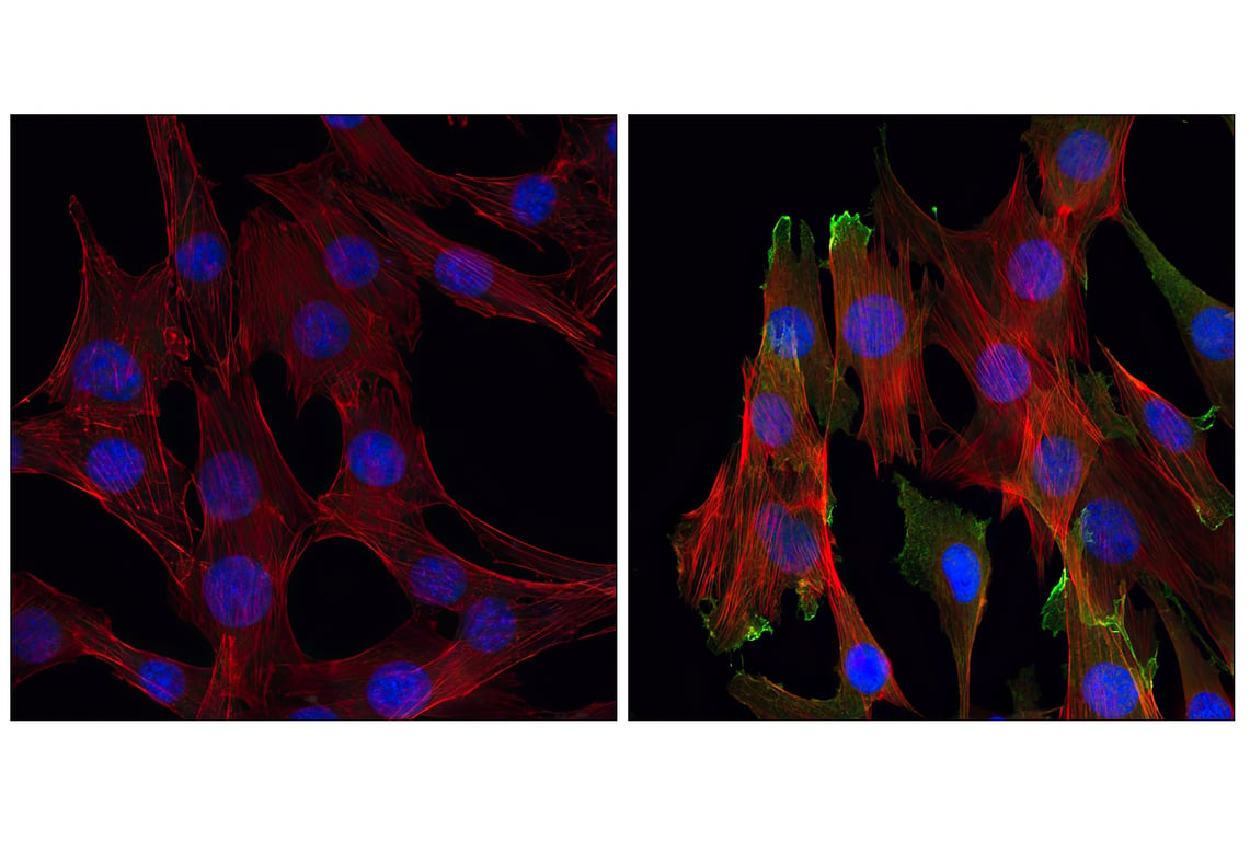
Confocal immunofluorescent analysis of C2C12 cells, LY294002-treated (left) or insulin-treated (right), using Phospho-Akt (Ser473) (D9E) XP® Rabbit mAb (green). Actin filaments have been labeled with Alexa Fluor® 555 phalloidin #8953 (red). Blue pseudocolor = DRAQ5®#4084 (fluorescent DNA dye).