


 全部商品分类


Akt (pan) (C67E7) Rabbit mAb


 下载产品说明书 下载COA 下载SDS
 用小程序,查商品更便捷


 收藏
收藏
 对比
对比 咨询
咨询
Monoclonal antibody is produced by immunizing animals with a synthetic peptide corresponding to residues in the carboxy-terminal sequence of mouse Akt.
Product Usage Information
| Application | Dilution |
|---|---|
| Western Blotting | 1:1000 |
| Immunoprecipitation | 1:50 |
| Immunohistochemistry (Paraffin) | 1:150 - 1:600 |
| Immunofluorescence (Immunocytochemistry) | 1:200 - 1:800 |
| Flow Cytometry (Fixed/Permeabilized) | 1:100 - 1:400 |
Specificity/Sensitivity
Species Reactivity:
Human, Mouse, Rat, Monkey, D. melanogaster
Supplied in 10 mM sodium HEPES (pH 7.5), 150 mM NaCl, 100 µg/ml BSA, 50% glycerol and less than 0.02% sodium azide. Store at –20°C. Do not aliquot the antibody.
For a carrier free (BSA and azide free) version of this product see product #88800.
参考图片
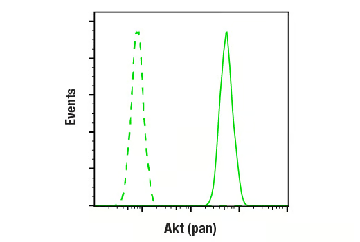
Flow cytometric analysis of Jurkat cells using Akt (pan) (C67E7) Rabbit mAb (solid line) compared to concentration-matched Rabbit (DA1E) mAb IgG XP® Isotype control #3900 (dashed line). Anti-rabbit IgG (H+L), F(ab')2 Fragment (Alexa Fluor® 488 Conjugate) #4412 was used as a secondary antibody.
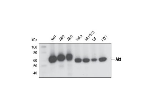
Western blot analysis of recombinant Akt1, Akt2 and Akt3 proteins, and extracts from various cell lines, using Akt (pan) (C67E7) Rabbit mAb.
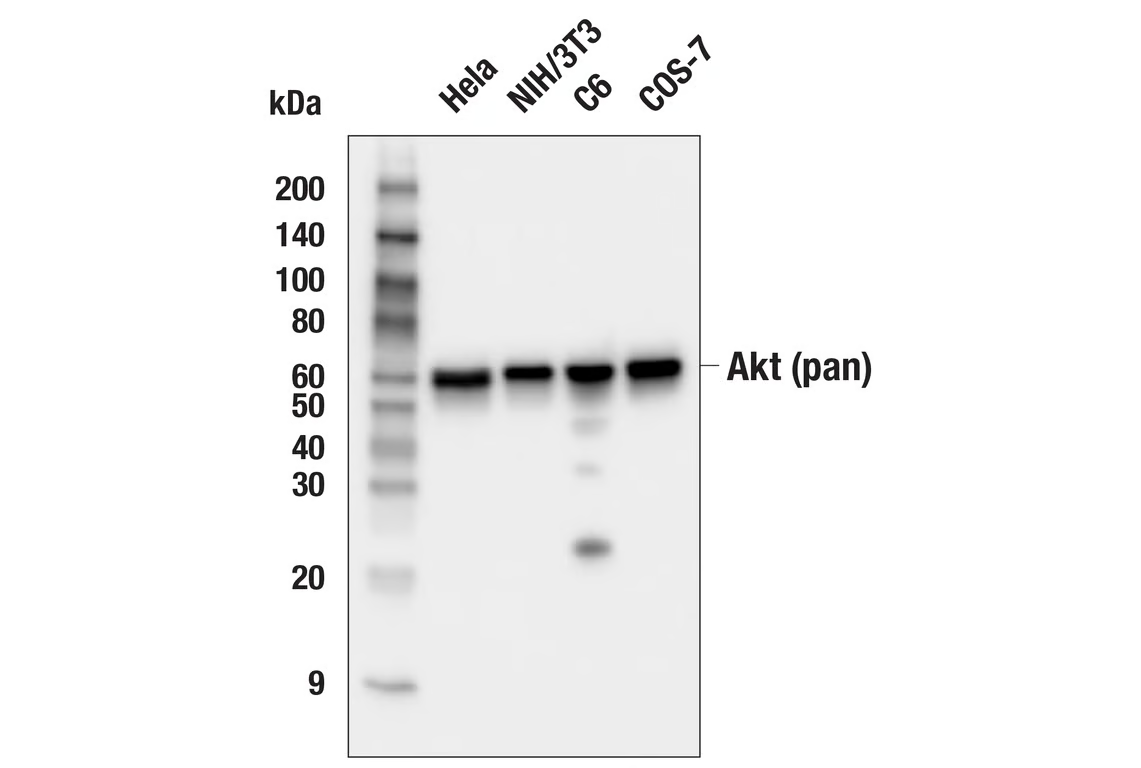
Western blot analysis of extracts from various cell lines using Akt (pan) (C67E7) Rabbit mAb #4691
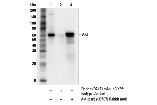
Immunoprecipitation of Akt from NIH/3T3 cell extracts. Lane 1 is 10% input, lane 2 is Rabbit (DA1E) mAb IgG XP® Isotype Control #3900, and lane 3 is Akt (pan) (C67E7) Rabbit mAb #4691. Western blot analysis was performed using Akt (pan) (40D4) Mouse mAb #2920.
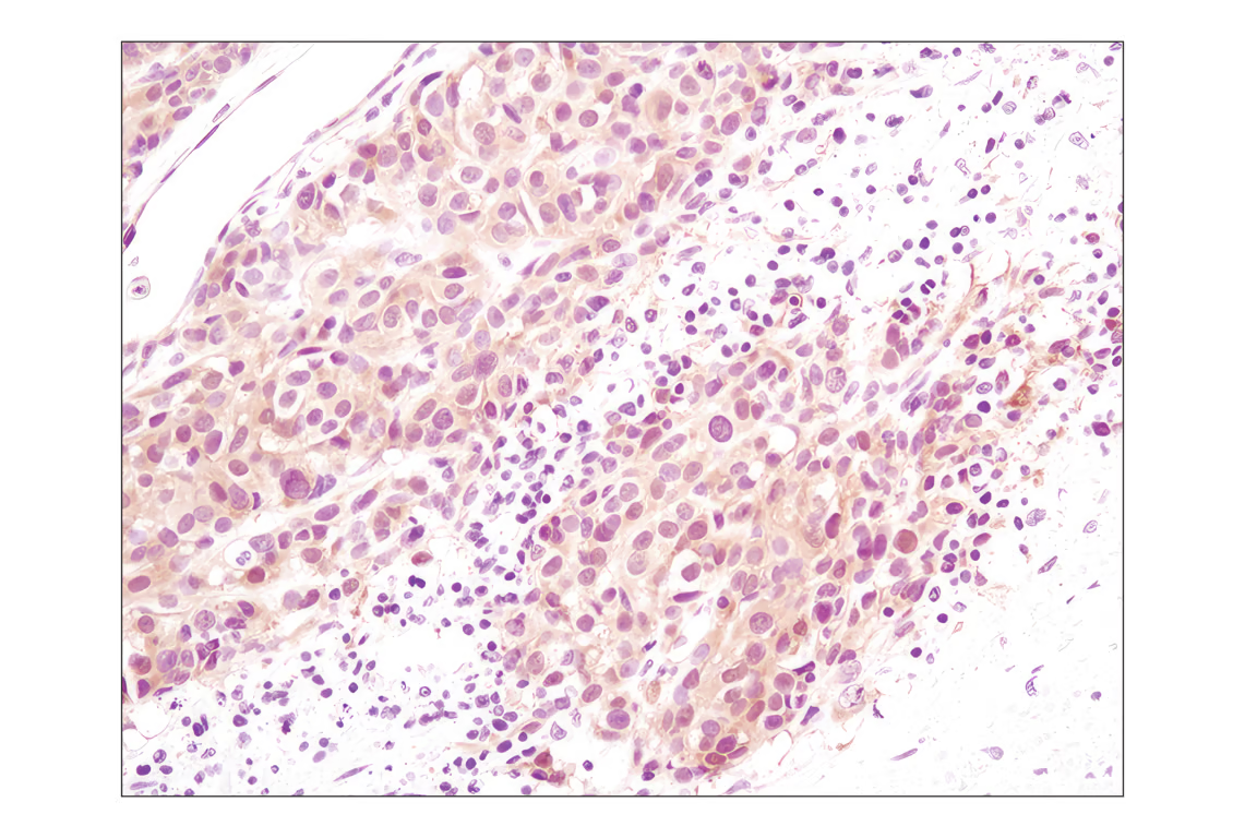
Immunohistochemical analysis of paraffin-embedded human melanoma using Akt (pan) (C67E7) Rabbit mAb.
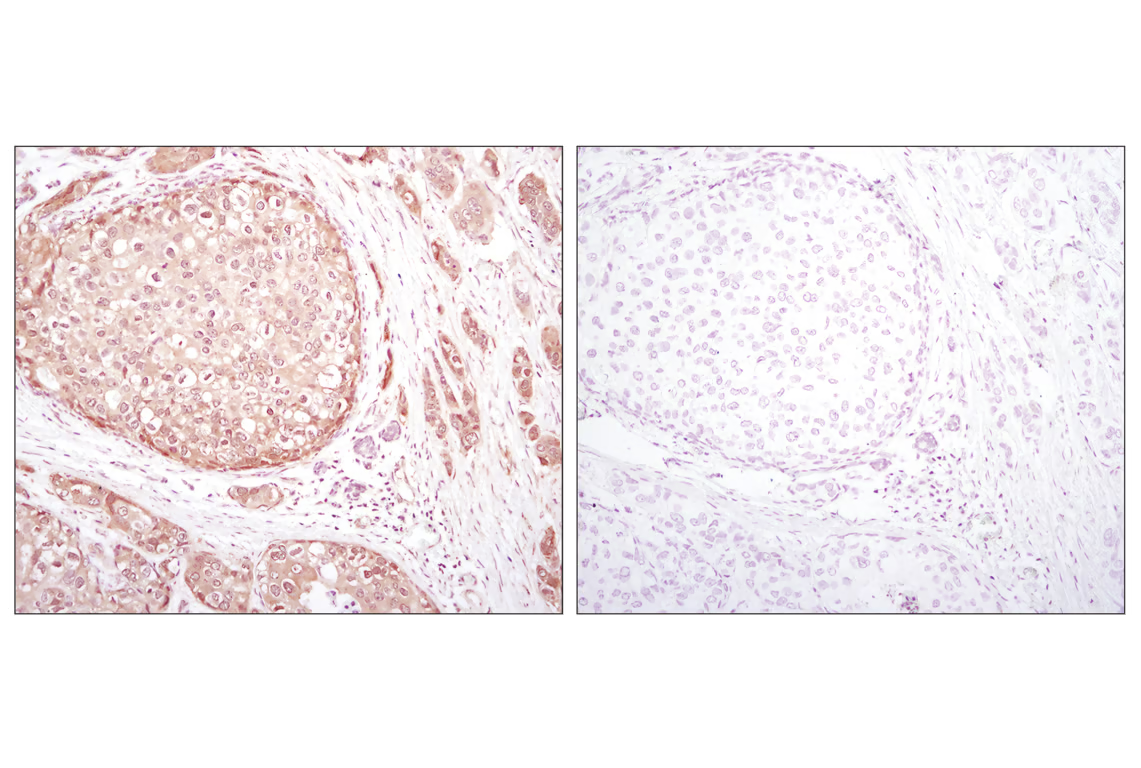
Immunohistochemical analysis of paraffin-embedded human breast carcinoma using Akt (pan) (C67E7) Rabbit mAb in the presence of control peptide (left) or Akt (pan) Blocking Peptide #1085 (right).
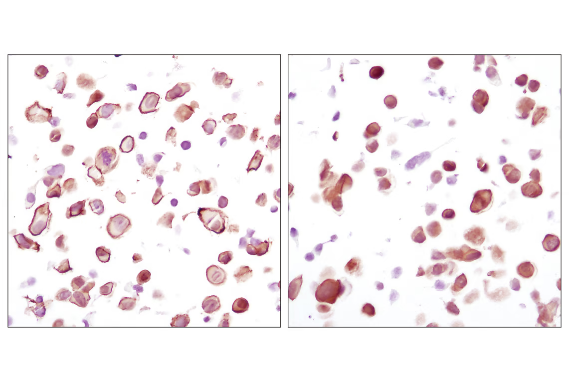
Immunohistochemical analysis using Akt (pan) (C67E7) Rabbit mAb on SignalSlide (TM) Phospho-Akt (Ser473) IHC Controls #8101 (paraffin-embedded LNCaP cells, untreated (left) or LY294002-treated (right)).
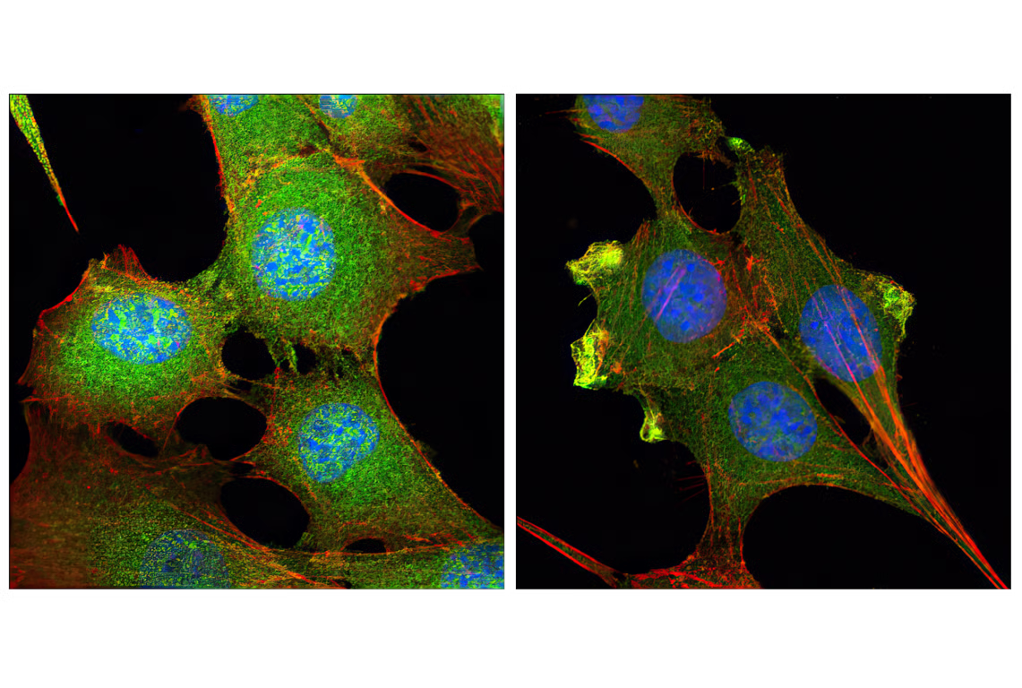
Confocal immunofluorescent analysis of C2C12 cells, LY294002-treated (left) or insulin-treated (right), using Akt (pan) (C67E7) Rabbit mAb (green). Actin filaments have been labeled with Alexa Fluor® 555 phalloidin (red). Blue pseudocolor = DRAQ5™ (fluorescent DNA dye).