


 全部商品分类
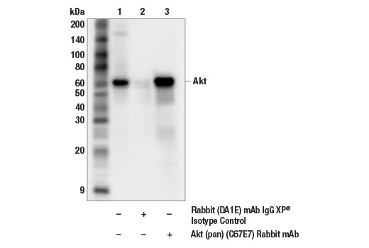


PhosphoPlus ® Akt (Ser473) Antibody Duet

 下载产品说明书 下载SDS
 用小程序,查商品更便捷


 收藏
收藏
 对比
对比 咨询
咨询
PhosphoPlus® Duets from Cell Signaling Technology (CST) provide a means to assess protein activation status. Each Duet contains an activation-state and total protein antibody to your target of interest. These antibodies have been selected from CST's product offering based upon superior performance in specified applications.
参考图片
Immunoprecipitation of Akt from NIH/3T3 cell extracts. Lane 1 is 10% input, lane 2 is Rabbit (DA1E) mAb IgG XP® Isotype Control #3900, and lane 3 is Akt (pan) (C67E7) Rabbit mAb #4691. Western blot analysis was performed using Akt (pan) (40D4) Mouse mAb #2920.
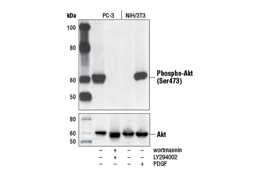
Western blot analysis of extracts from PC-3 cells, untreated or LY294002/wortmannin-treated, and NIH/3T3 cells, serum-starved or PDGF-treated, using Phospho-Akt (Ser473) (D9E) XP® Rabbit mAb (upper) or Akt (pan) (C67E7) Rabbit mAb #4691 (lower).
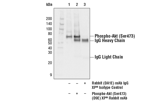
Immunoprecipitation of phospho-Akt (Ser473) from Jurkat extracts treated with Calyculin A #9902 (100nM, 30 min). Lane 1 is 10% input, lane 2 is Phospho-Akt (Ser473) (D9E) XP® Rabbit mAb, and lane 3 is Rabbit (DA1E) mAb IgG XP® Isotype Control #3900. Western blot analysis was performed with Phospho-Akt (Ser473) (D9E) XP® Rabbit mAb. Anti-rabbit IgG, HRP-linked Antibody #7074 was used as a secondary antibody.
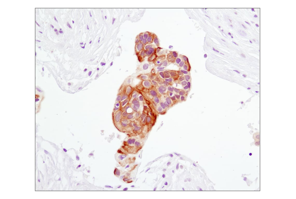
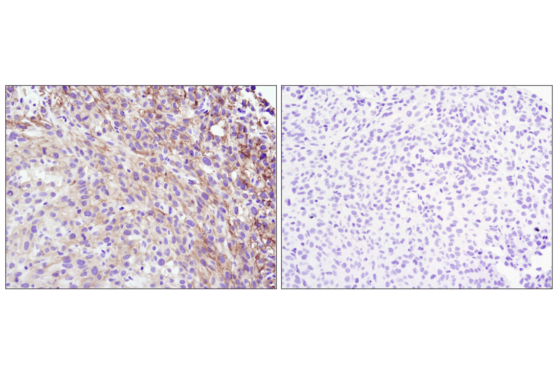
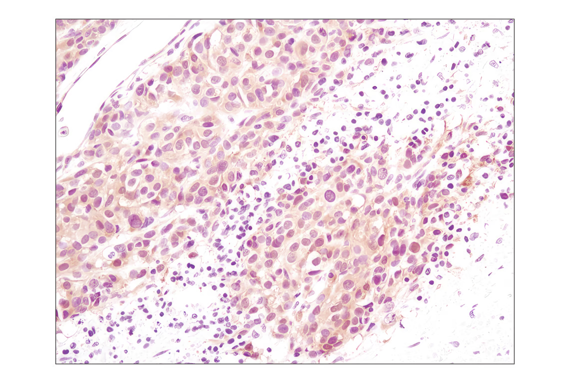
Immunohistochemical analysis of paraffin-embedded human lung carcinoma using Phospho-Akt (Ser473) (D9E) XP® Rabbit mAb.
Immunohistochemical analysis of paraffin-embedded human breast carcinoma using Phospho-Akt (Ser473) (D9E) XP® Rabbit mAb.
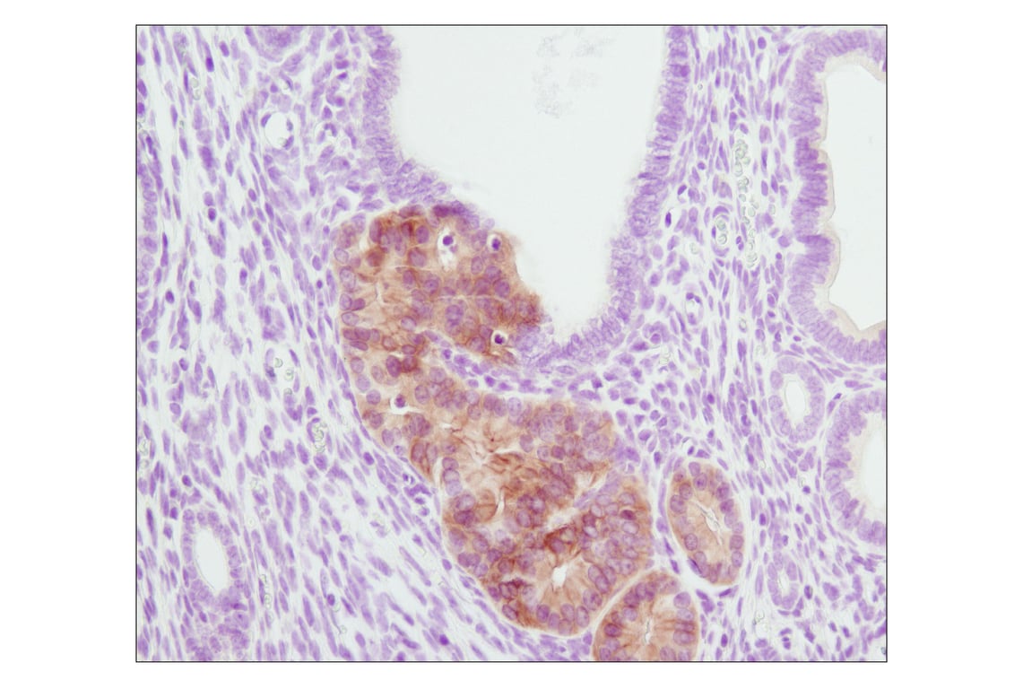
Immunohistochemical analysis of paraffin-embedded PTEN heterozygous mutant mouse endometrium using Phospho-Akt (Ser473) (D9E) XP® Rabbit mAb. (Tissue section courtesy of Dr. Sabina Signoretti, Brigham and Women's Hospital, Harvard Medical School, Boston, MA.)
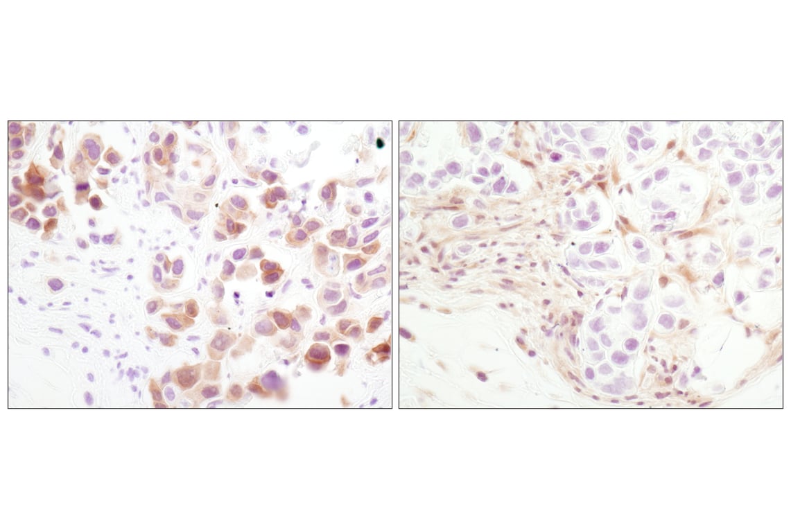
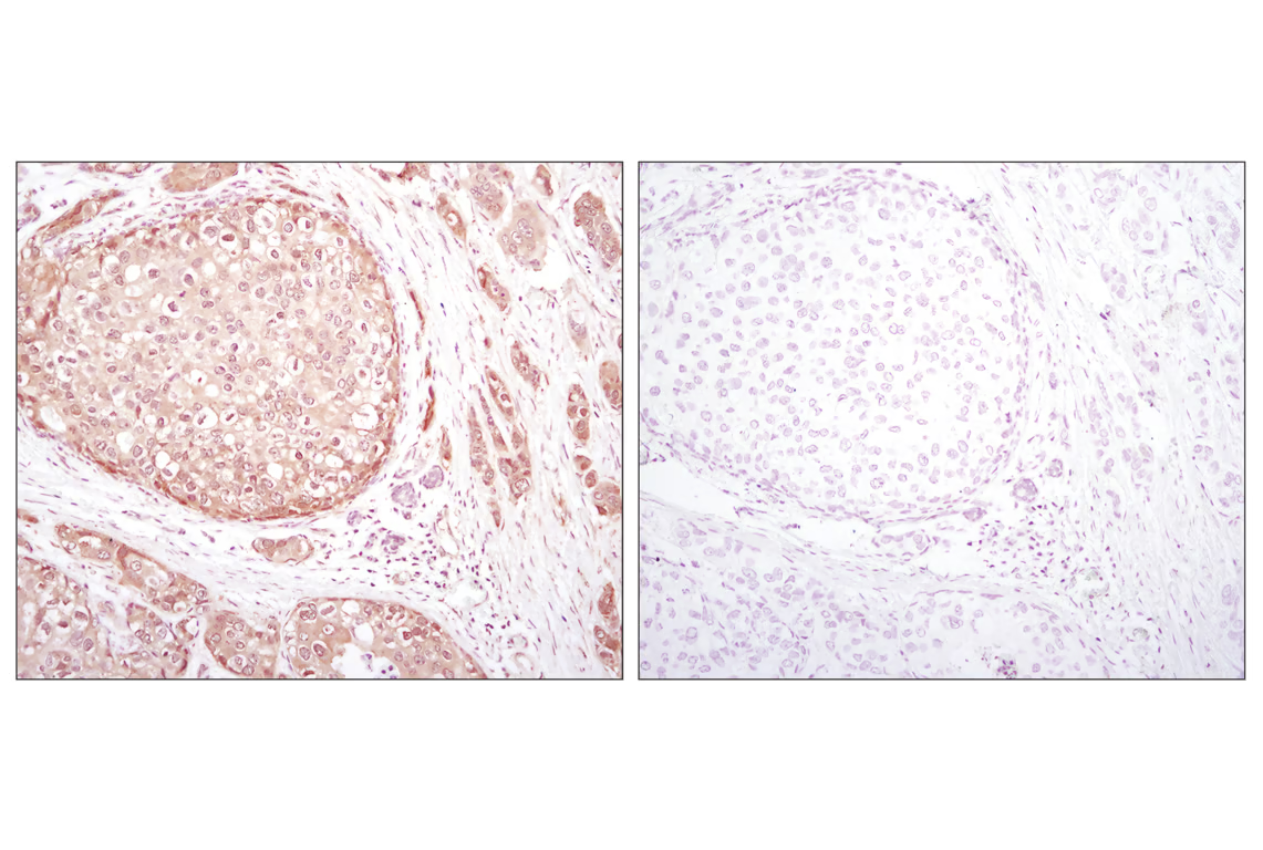
Immunohistochemical analysis of paraffin-embedded MDA-MB-468 xenograft using Phospho-Akt (Ser473) (D9E) XP® Rabbit mAb (left) or PTEN (138G6) Rabbit mAb #9559 (right). Note the presence of P-Akt staining in the PTEN deficient MDA-MB-468 cells.
Immunohistochemical analysis of paraffin-embedded human breast carcinoma comparing SignalStain® Antibody Diluent #8112 (left) to TBST/5% normal goat serum (right) using Phospho-Akt (Ser473) (D9E) XP® Rabbit mAb #4060.
Immunohistochemical analysis of paraffin-embedded U-87MG xenograft, untreated (left) or lambda phosphatase-treated (right), using Phospho-Akt (Ser473) (D9E) XP® Rabbit mAb.
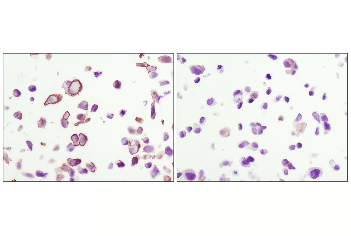
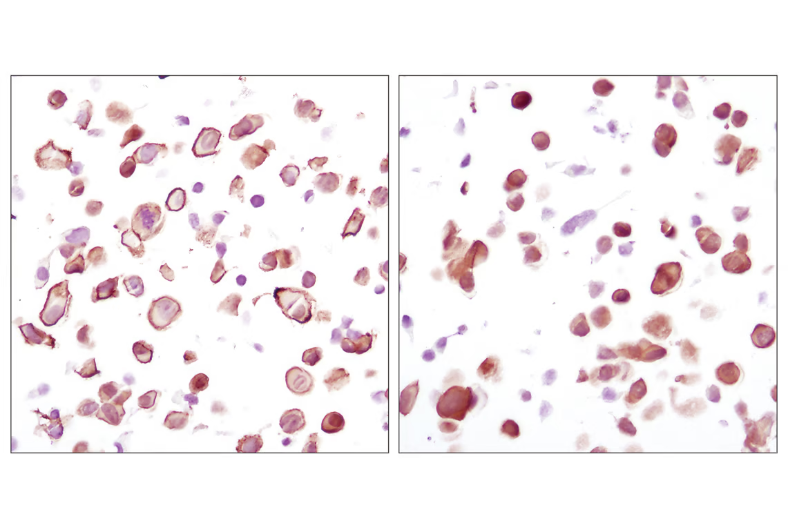
Immunohistochemical analysis using Phospho-Akt (Ser473) (D9E) XP® Rabbit mAb on SignalSlide® Phospho-Akt (Ser473) IHC Controls #8101 (paraffin-embedded LNCaP cells, untreated (left) or LY294002-treated (right)).
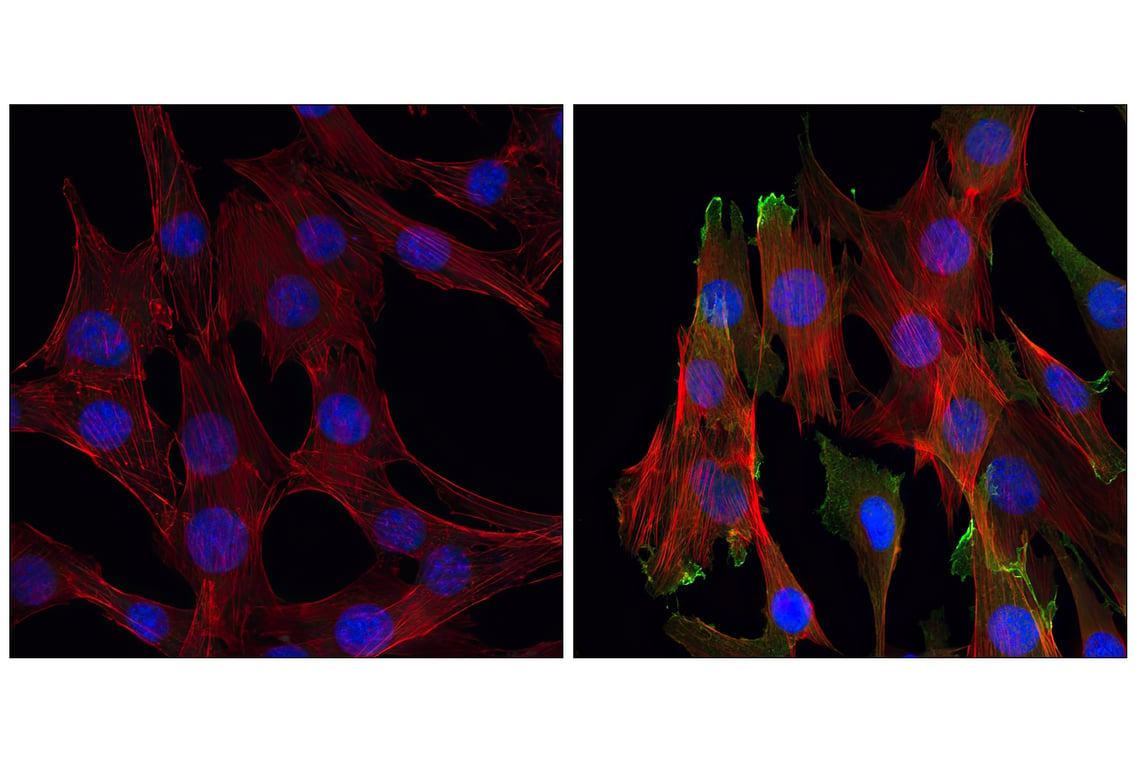
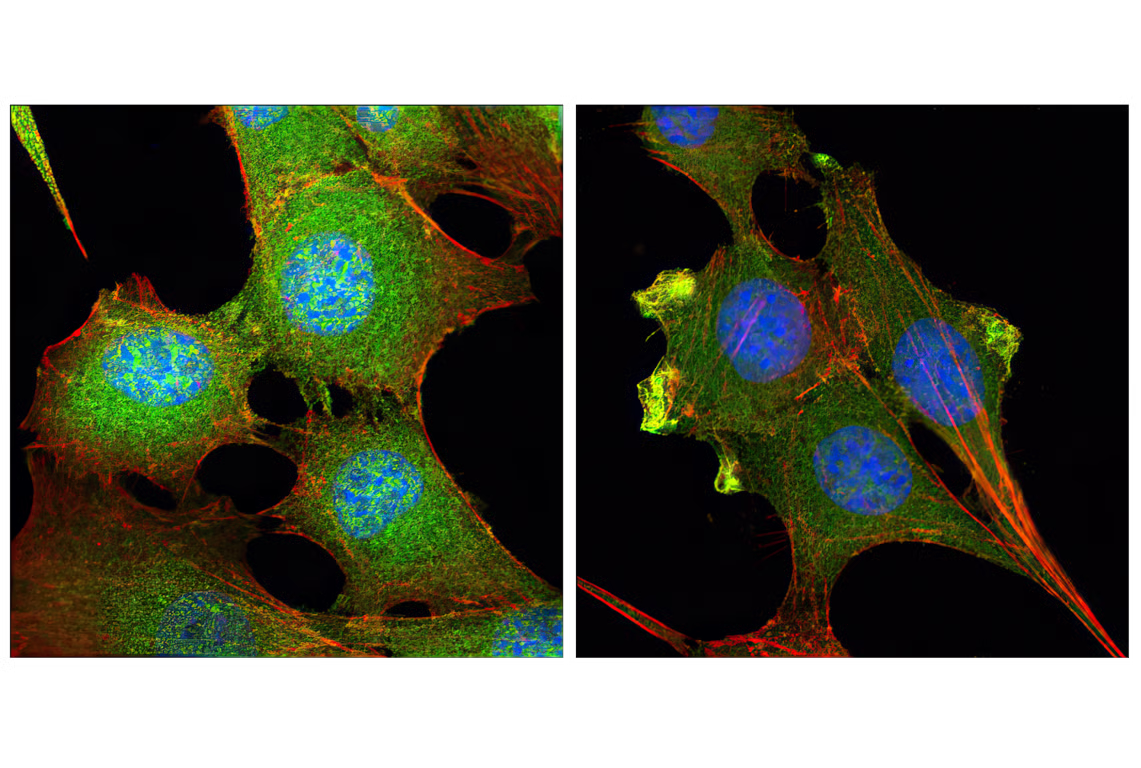
Confocal immunofluorescent analysis of C2C12 cells, LY294002-treated (left) or insulin-treated (right), using Phospho-Akt (Ser473) (D9E) XP® Rabbit mAb (green). Actin filaments have been labeled with Alexa Fluor® 555 phalloidin #8953 (red). Blue pseudocolor = DRAQ5®#4084 (fluorescent DNA dye).
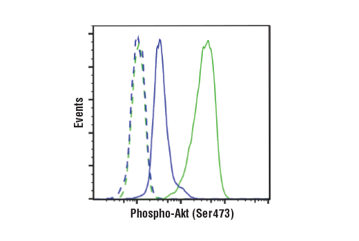
Flow cytometric analysis of Jurkat cells, untreated (green) or treated with LY294002 #9901, Wortmannin #9951, and U0126 #9903 (50 μM, 1 μM, and 10 μM, 2 hr; blue) using Phospho-Akt (Ser473) (D9E) XP® Rabbit mAb (solid lines) or concentration-matched Rabbit (DA1E) mAb IgG XP® Isotype Control #3900 (dashed lines). Anti-rabbit IgG (H+L), F(ab')2 Fragment (Alexa Fluor® 488 Conjugate) #4412 was used as a secondary antibody.
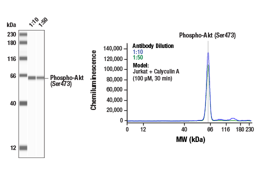
Simple Western™ analysis of lysates (0.1 mg/mL) from Jurkat cells treated with Calyculin A (100 uM, 30 min) using Phospho-Akt (Ser473) (D9E) XP® Rabbit mAb #4060. The virtual lane view (left) shows a single target band (as indicated) at 1:10 and 1:50 dilutions of primary antibody. The corresponding electropherogram view (right) plots chemiluminescence by molecular weight along the capillary at 1:10 (blue line) and 1:50 (green line) dilutions of primary antibody. This experiment was performed under reducing conditions on the Jess™ Simple Western instrument from ProteinSimple, a BioTechne brand, using the 12-230 kDa separation module.
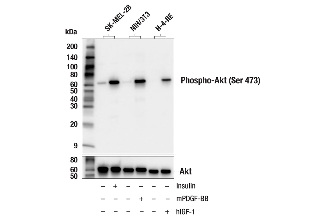
Western blot analysis of extracts from various cell lines, untreated (-) or treated (+) as indicated with human insulin (100 nM, 20 min), mouse PDGF-BB (100 ng/ml, 20 min), or human Insulin-like Growth Factor I (hIGF-I) #8917 (100 ng/ml; 5 min), using Phospho-Akt (Ser473) (D9E) XP® Rabbit mAb (upper) or Akt (pan) (C67E7) Rabbit mAb #4691 (lower).
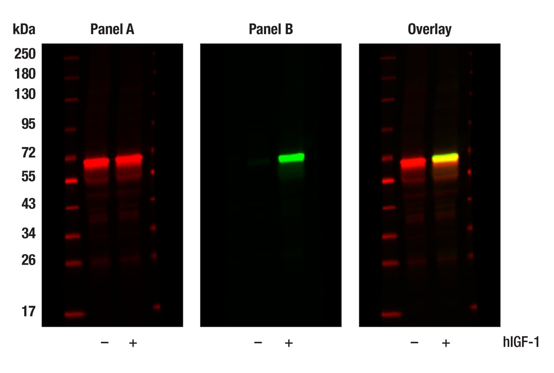
Western blot analysis of extracts from MCF-7 cells, untreated (-) or treated with hIGF-1 (100 ng/ml, 10 min; +), using Akt (pan) (E7J2C) Mouse mAb #58295 (Panel A) and Phospho-Akt (Ser473) (D9E) XP® Rabbit mAb #4060 (Panel B). Anti-mouse IgG (H+L) (DyLight 680 Conjugate) #5470 (red) and Anti-rabbit IgG (H+L) (DyLight 800 4X PEG Conjugate) #5151 (green) were used as secondary antibodies.
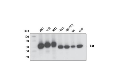
Western blot analysis of recombinant Akt1, Akt2 and Akt3 proteins, and extracts from various cell lines, using Akt (pan) (C67E7) Rabbit mAb.
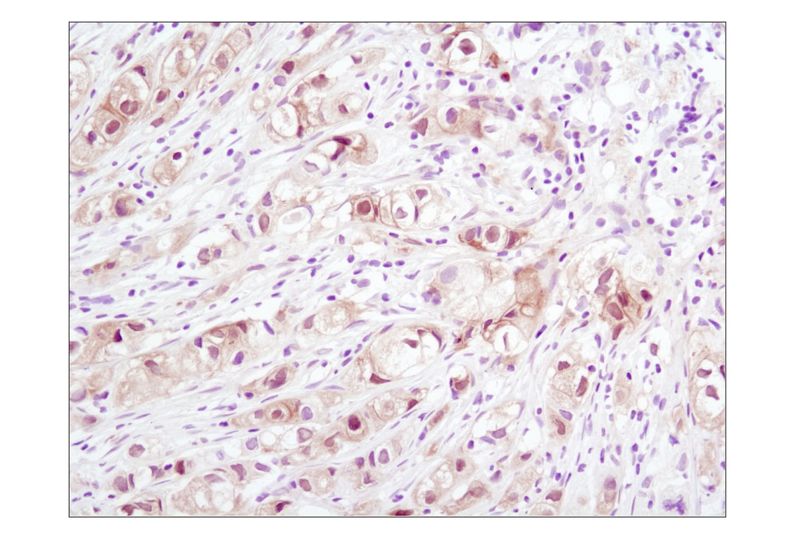
Immunohistochemical analysis of paraffin-embedded human melanoma using Akt (pan) (C67E7) Rabbit mAb.
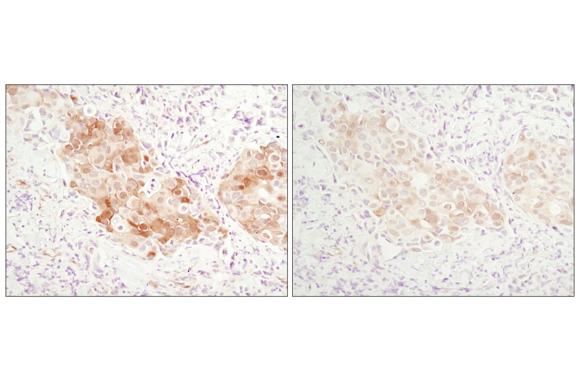
Immunohistochemical analysis of paraffin-embedded human breast carcinoma using Akt (pan) (C67E7) Rabbit mAb in the presence of control peptide (left) or Akt (pan) Blocking Peptide #1085 (right).
Immunohistochemical analysis using Akt (pan) (C67E7) Rabbit mAb on SignalSlide (TM) Phospho-Akt (Ser473) IHC Controls #8101 (paraffin-embedded LNCaP cells, untreated (left) or LY294002-treated (right)).
Confocal immunofluorescent analysis of C2C12 cells, LY294002-treated (left) or insulin-treated (right), using Akt (pan) (C67E7) Rabbit mAb (green). Actin filaments have been labeled with Alexa Fluor® 555 phalloidin (red). Blue pseudocolor = DRAQ5™ (fluorescent DNA dye).
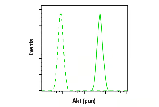
Flow cytometric analysis of Jurkat cells using Akt (pan) (C67E7) Rabbit mAb (solid line) compared to concentration-matched Rabbit (DA1E) mAb IgG XP® Isotype control #3900 (dashed line). Anti-rabbit IgG (H+L), F(ab')2 Fragment (Alexa Fluor® 488 Conjugate) #4412 was used as a secondary antibody.
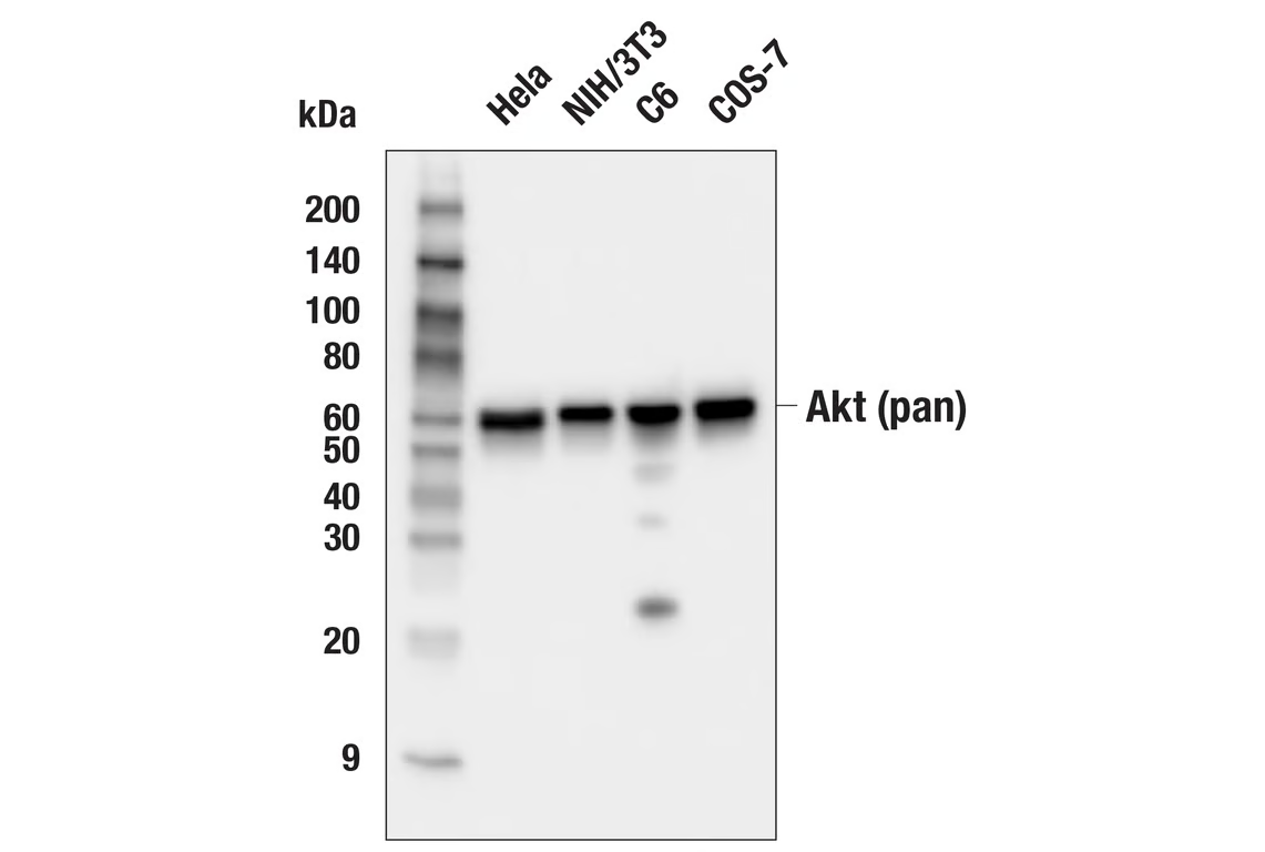
Western blot analysis of extracts from various cell lines using Akt (pan) (C67E7) Rabbit mAb #4691