


 全部商品分类
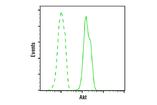


Akt Antibody


 下载产品说明书 下载COA 下载SDS
 用小程序,查商品更便捷


 收藏
收藏
 对比
对比 咨询
咨询
Polyclonal antibodies are produced by immunizing animals with a synthetic peptide corresponding to the carboxy-terminal sequence of mouse Akt. Antibodies are purified by protein A and peptide affinity chromatography.
Product Usage Information
| Application | Dilution |
|---|---|
| Western Blotting | 1:1000 |
| Immunoprecipitation | 1:50 |
| Immunofluorescence (Immunocytochemistry) | 1:200 |
| Flow Cytometry (Fixed/Permeabilized) | 1:50 - 1:200 |
Specificity/Sensitivity
Species Reactivity:
Human, Mouse, Rat, Hamster, Monkey, Chicken, D. melanogaster, Bovine, Dog, Pig, Guinea Pig
Supplied in 10 mM sodium HEPES (pH 7.5), 150 mM NaCl, 100 µg/ml BSA and 50% glycerol. Store at –20°C. Do not aliquot the antibody.
参考图片
Flow cytometric analysis of Jurkat cells using Akt Antibody (solid line) compared to concentration-matched Rabbit (DA1E) mAb IgG XP® Isotype Control #3900 (dashed line). Anti-rabbit IgG (H+L), F(ab')2 Fragment (Alexa Fluor® 488 Conjugate) #4412 was used as a secondary antibody.
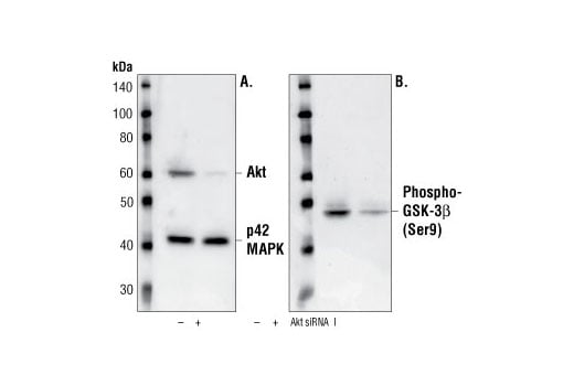
Western blot analysis of extracts from CHO cells, transfected with non-targeted (-) or SignalSilence® Akt siRNA I (+) siRNA, using Akt Antibody #9272 and p42 MAP Kinase (Erk2) Antibody #9108. The Akt antibody confirms silencing of protein expression while the p42 MAP Kinase (Erk2) antibody was used to control for loading and specificity of Akt siRNA (A). Phospho-GSK-3β (Ser9) Antibody #9336 was used to confirm downstream pathway inhibition (B).
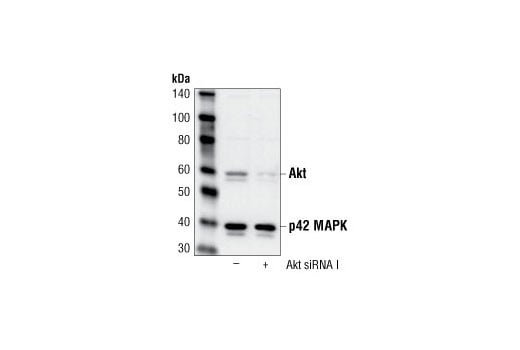
Western blot analysis of extracts from HeLa cells, transfected with 100 nM SignalSilence® Control siRNA (Fluorescein Conjugate) #6201 (-) or SignalSilence® Akt siRNA I (+), using Akt Antibody #9272 and p42 MAP Kinase (Erk2) Antibody #9108. Akt antibody confirms silencing of Akt expression, while the p42 MAP kinase (Erk2) antibody is used to control for loading and specificity of Akt siRNA.
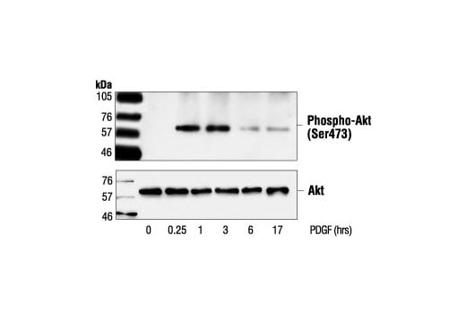
Western blot analysis of extracts from NIH/3T3 cells, untreated or PDGF-treated (50 ng/ml) for the indicated times, using Phospho-Akt (Ser473) Antibody #9271 (upper) or Akt Antibody (lower).
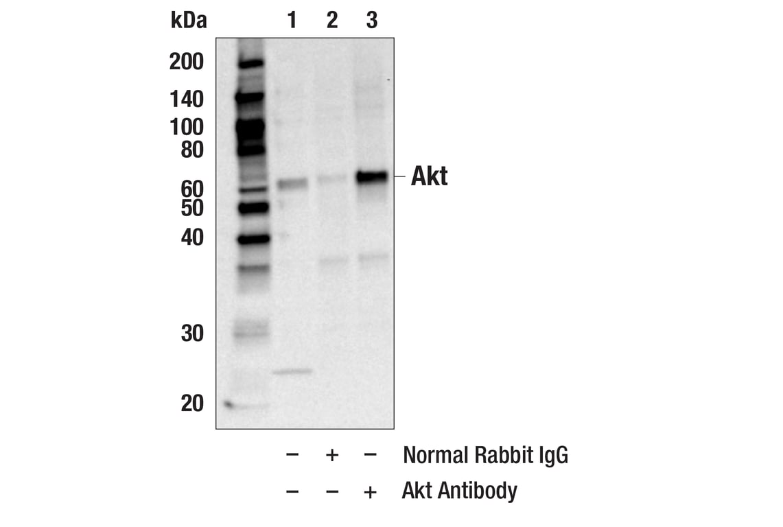
Immunoprecipitation of Akt from HeLa cell extracts. Lane 1 is 10% input, lane 2 is Normal Rabbit IgG #2729, and lane 3 is Akt Antibody. Western blot was performed using Akt Antibody. Mouse Anti-rabbit IgG (Conformation Specific) (L27A9) mAb #3678 was used as a secondary antibody.
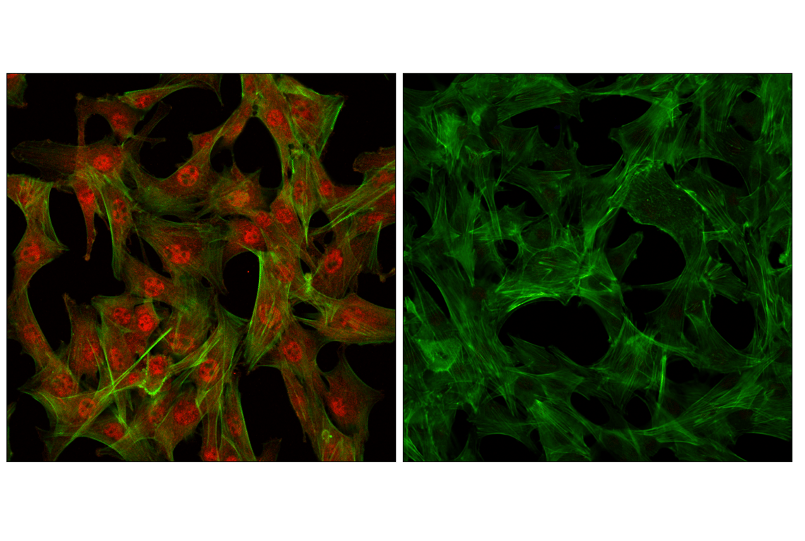
Confocal immunofluorescent images of C2C12 cells showing nuclear and cytoplasmic localization with Akt Antibody (left, red) compared to an isotype control (right). Actin filaments have been labeled with fluorescein phalloidin.