


 全部商品分类


Phospho-PRAS40 (Thr246) (C77D7) Rabbit mAb

 下载产品说明书 下载COA 下载SDS
 用小程序,查商品更便捷


 收藏
收藏
 对比
对比 咨询
咨询
Monoclonal antibody is produced by immunizing animals with a synthetic phosphopeptide corresponding to the sequence surrounding Thr246 of human PRAS40.
Product Usage Information
| Application | Dilution |
|---|---|
| Western Blotting | 1:1000 |
| Simple Western™ | 1:10 - 1:50 |
| Immunoprecipitation | 1:50 |
| Immunohistochemistry (Paraffin) | 1:800 - 1:3200 |
Specificity/Sensitivity
Species Reactivity:
Human, Mouse, Rat, Monkey
Supplied in 10 mM sodium HEPES (pH 7.5), 150 mM NaCl, 100 µg/ml BSA, 50% glycerol and less than 0.02% sodium azide. Store at –20°C. Do not aliquot the antibody.
For a carrier free (BSA and azide free) version of this product see product #60202.
参考图片
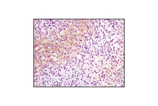
Immunohistochemical analysis of paraffin-embedded metastatic SKOV-3 tumor in mouse lung using Phospho-PRAS40 (Thr246) (C77D7) Rabbit mAb.
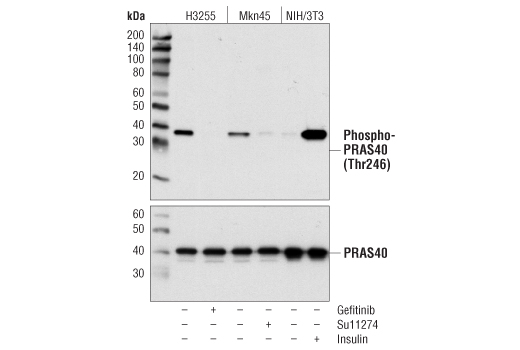
Western blot analysis of extracts from serum starved H3255, Mkn45 and NIH/3T3 cells, untreated or treated with either Gefitinib (1 μM, 3 hours), Su11274 (1 μM, 3 hours) or insulin (150 nM, 15 minutes), using Phospho-PRAS40 (Thr246) (C77D7) Rabbit mAb (upper) or PRAS40 (D23C7) Rabbit mAb #2691 (lower).
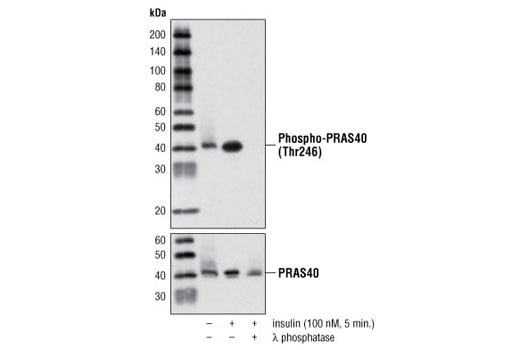
Western blot analysis of extracts from serum starved HeLa cells, untreated or treated with insulin (100 nM, 5 minutes) or with insulin and λ phosphatase, using Phospho-PRAS40 (Thr246) (C77D7) Rabbit mAb (upper) or PRAS40 Antibody #2610 (lower).
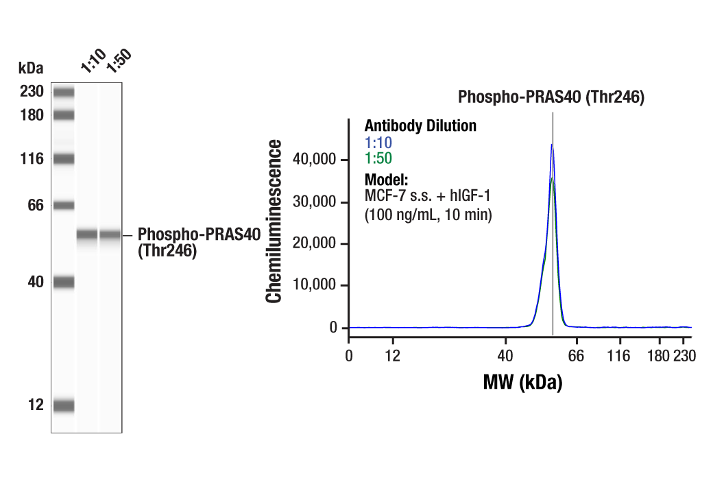
Simple Western™ analysis of lysates (1.0 mg/mL) from serum-starved MCF-7 cells treated with human IGF-1 (100 ng/mL, 10 min) using Phospho-PRAS40 (Thr246) (C77D7) Rabbit mAb #2997. The virtual lane view (left) shows a single target band (as indicated) at 1:10 and 1:50 dilutions of primary antibody. The corresponding electropherogram view (right) plots chemiluminescence by molecular weight along the capillary at 1:10 (blue line) and 1:50 (green line) dilutions of primary antibody. This experiment was performed under reducing conditions on the Jess™ Simple Western instrument from ProteinSimple, a BioTechne brand, using the 12-230 kDa separation module.
Immunoprecipitation of Phospho-PRAS40 (Thr246) from MCF-7 cell extracts. Cells were treated with hIGF-I (100 ng/mL, 10 min.). Lane 1 is 10% input, lane 2 is Rabbit (DA1E) mAb IgG XP® Isotype Control #3900, and lane 3 is Phospho-PRAS40 (Thr246) (C77D7) Rabbit mAb. Western blot analysis was performed using Phospho-PRAS40 (Thr246) (C77D7) Rabbit mAb. Anti-rabbit IgG, HRP-linked Antibody #7074 was used as a secondary antibody.
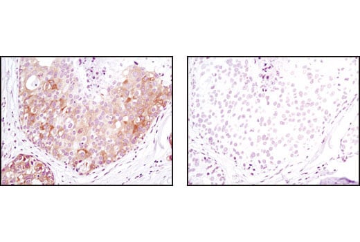
Immunohistochemical analysis of paraffin-embedded human breast carcinoma using Phospho-PRAS40 (Thr246) (C77D7) Rabbit mAb in the presence of control peptide (left) or antigen specific peptide (right).
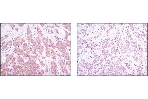
Immunohistochemical analysis of paraffin-embedded human breast carcinoma, control (left) or λ phosphatase-treated (right), using Phospho-PRAS40 (Thr246) (C77D7) Rabbit mAb.