


 全部商品分类
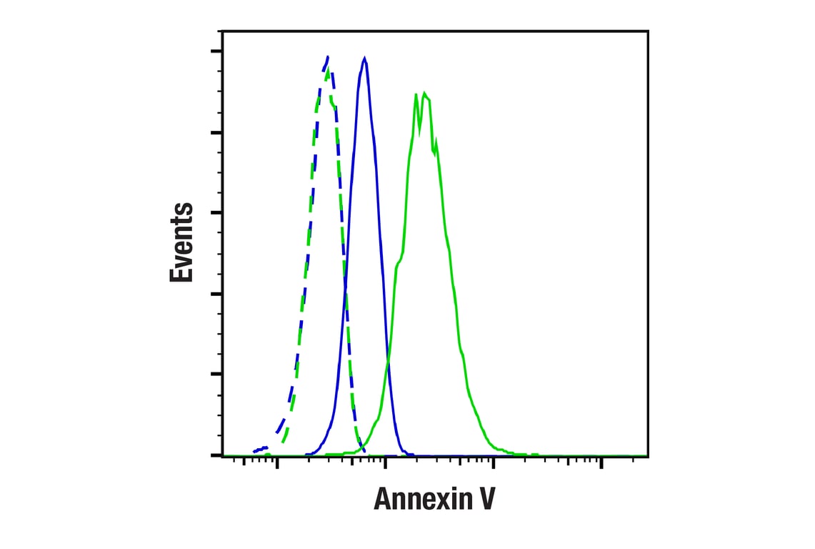


 用小程序,查商品更便捷


Monoclonal antibody is produced by immunizing animals with a synthetic peptide corresponding to residues surrounding Thr10 of human annexin V protein.

Product Usage Information
| Application | Dilution |
|---|---|
| Western Blotting | 1:1000 |
| Immunohistochemistry (Paraffin) | 1:50 - 1:200 |
| Immunofluorescence (Immunocytochemistry) | 1:100 - 1:400 |
| Flow Cytometry (Fixed/Permeabilized) | 1:100 - 1:400 |
Specificity/Sensitivity
Species Reactivity:
Human, Mouse, Rat
Supplied in 10 mM sodium HEPES (pH 7.5), 150 mM NaCl, 100 µg/mL BSA, 50% glycerol, and less than 0.02% sodium azide. Store at –20°C. Do not aliquot the antibody.
For a carrier free (BSA and azide free) version of this product see product #24122.
参考图片
Flow cytometric analysis of A20 cells (blue, low expressing) and NIH/3T3 cells (green, high expressing) using Annexin V (E3W8V) Rabbit mAb (solid lines) or concentration-matched Rabbit (DA1E) mAb IgG XP® Isotype Control #3900 (dashed lines). Anti-rabbit IgG (H+L), F(ab')2 Fragment (Alexa Fluor® 488 Conjugate) #4412 was used as a secondary antibody.
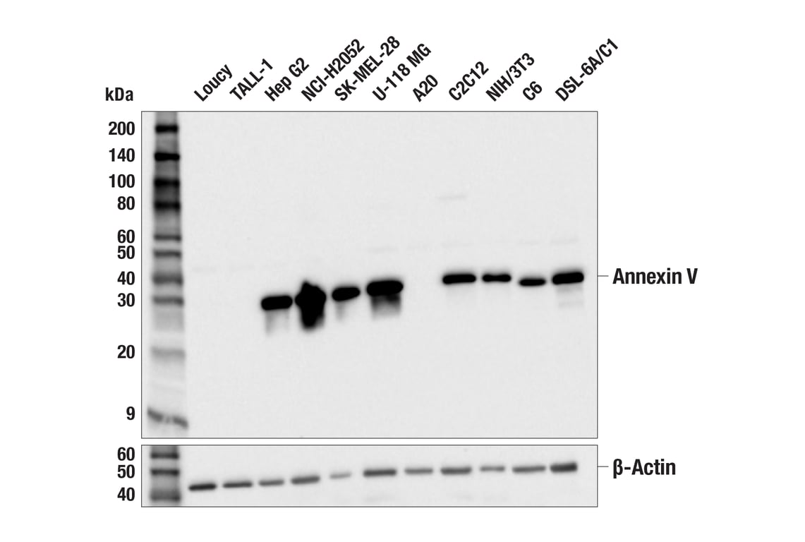
Western blot analysis of extracts from various cell lines using Annexin V (E3W8V) Rabbit mAb (upper) or β-Actin (D6A8) Rabbit mAb #8457 (lower). Negative expression of Annexin V protein in Loucy, TALL-1, and A20 cells is consistent with the predicted expression pattern.
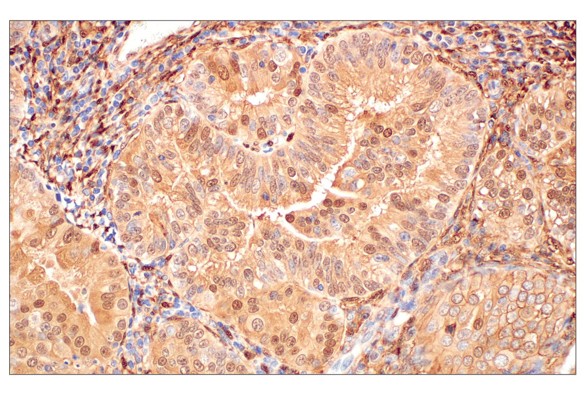
Immunohistochemical analysis of paraffin-embedded human endometrioid adenocarcinoma using Annexin V (E3W8V) Rabbit mAb.
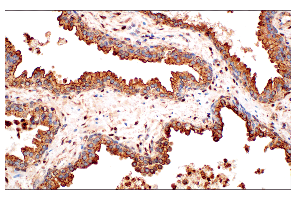
Immunohistochemical analysis of paraffin-embedded human squamous cell lung carcinoma using Annexin V (E3W8V) Rabbit mAb.
Immunohistochemical analysis of paraffin-embedded human prostate adenocarcinoma using Annexin V (E3W8V) Rabbit mAb.
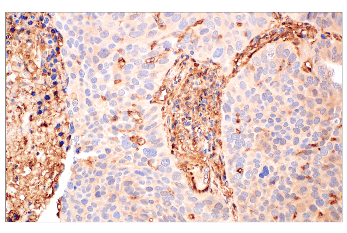
Immunohistochemical analysis of paraffin-embedded human urothelial carcinoma using Annexin V (E3W8V) Rabbit mAb.
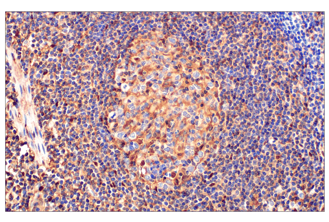
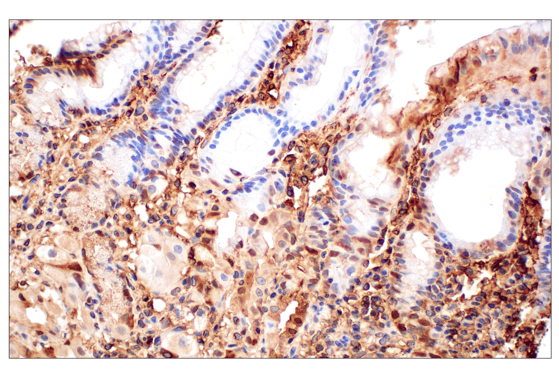
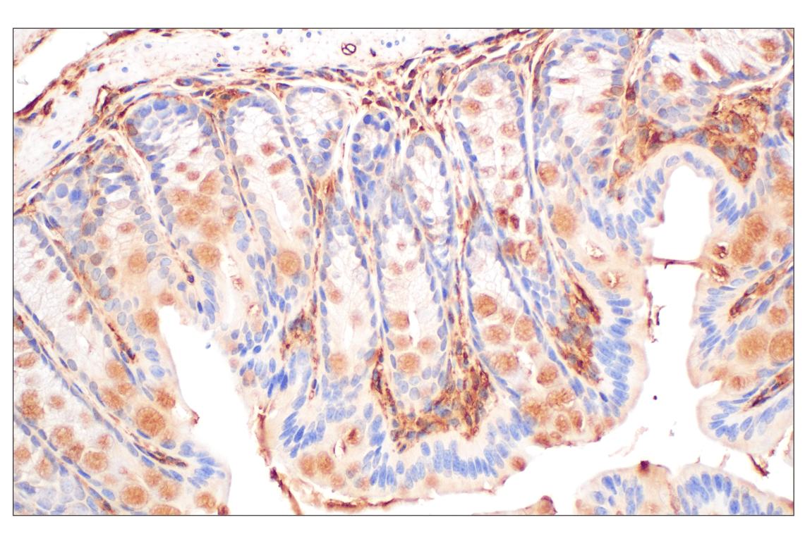
Immunohistochemical analysis of a Peyer's patch within paraffin-embedded normal human small intestine using Annexin V (E3W8V) Rabbit mAb.
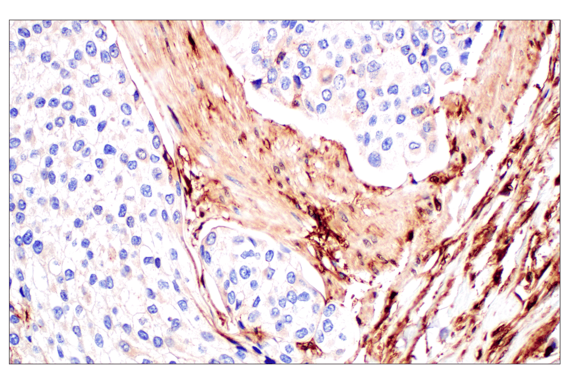
Immunohistochemical analysis of paraffin-embedded normal human liver using Annexin V (E3W8V) Rabbit mAb.
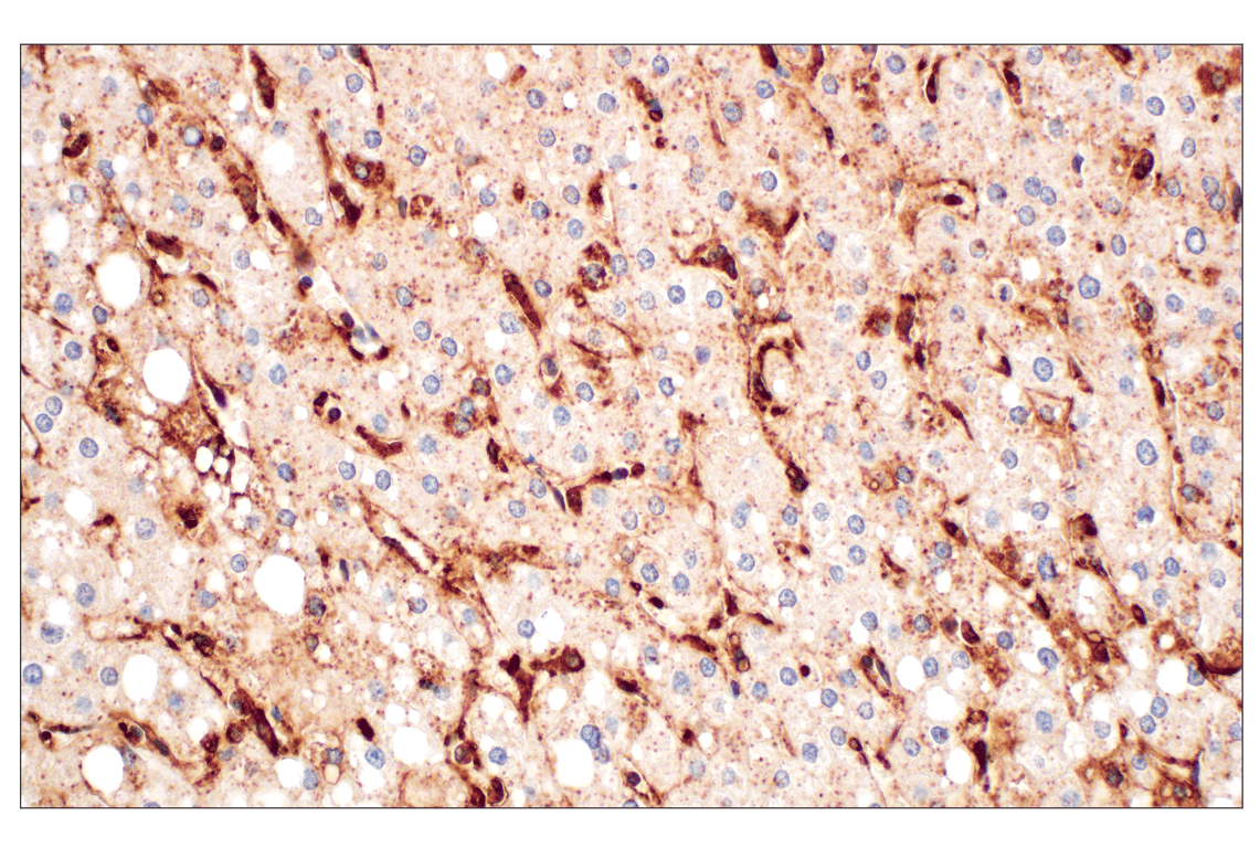
Immunohistochemical analysis of paraffin-embedded normal human stomach using Annexin V (E3W8V) Rabbit mAb.
Immunohistochemical analysis of paraffin-embedded 4T1 syngeneic mammary tumor using Annexin V (E3W8V) Rabbit mAb.
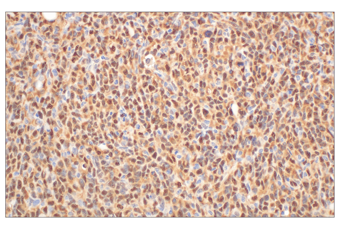
Immunohistochemical analysis of paraffin-embedded A20 syngeneic tumor using Annexin V (E3W8V) Rabbit mAb.
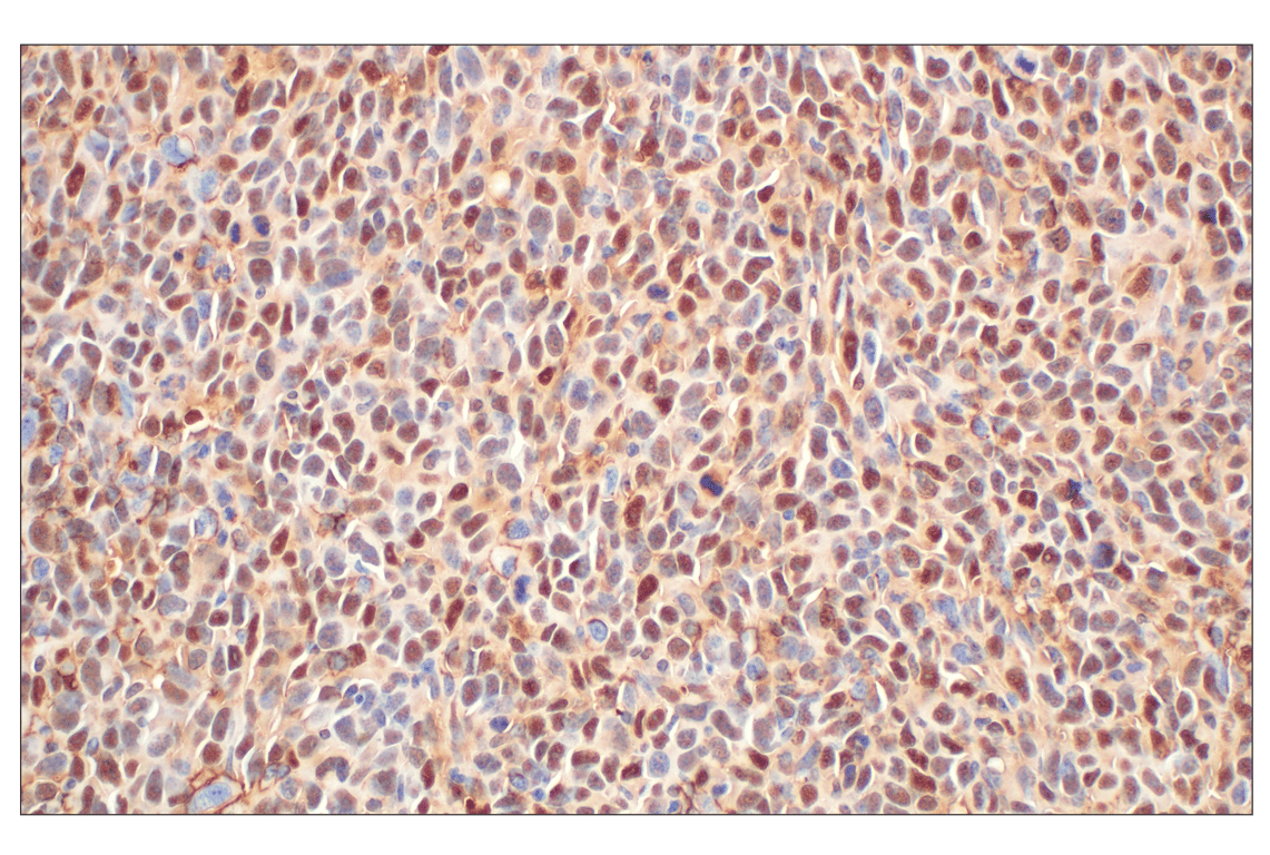
Immunohistochemical analysis of paraffin-embedded LL/2 syngeneic tumor using Annexin V (E3W8V) Rabbit mAb.
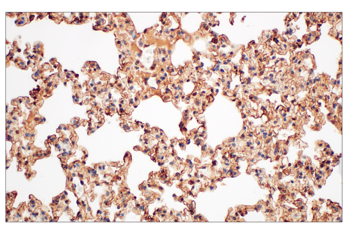
Immunohistochemical analysis of paraffin-embedded mouse lung using Annexin V (E3W8V) Rabbit mAb.
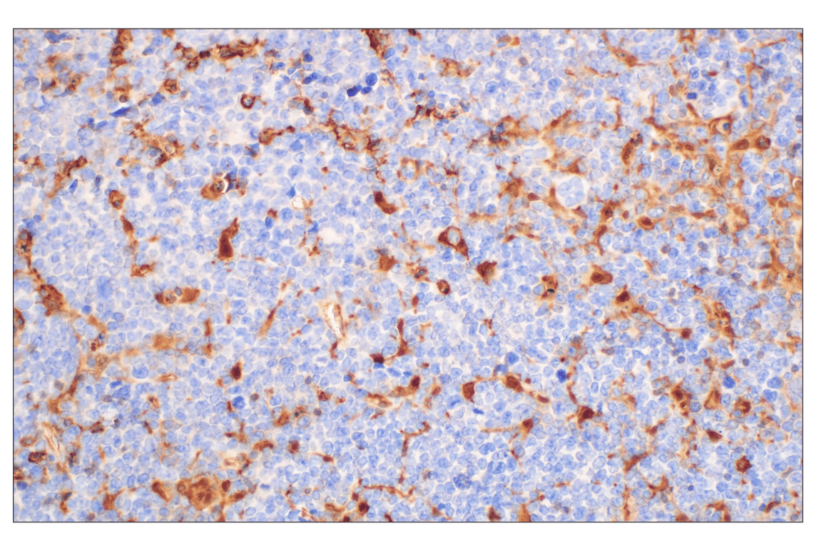
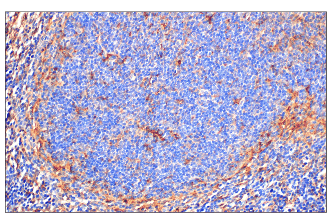
Immunohistochemical analysis of paraffin-embedded mouse spleen using Annexin V (E3W8V) Rabbit mAb.
Immunohistochemical analysis of paraffin-embedded mouse colon using Annexin V (E3W8V) Rabbit mAb.
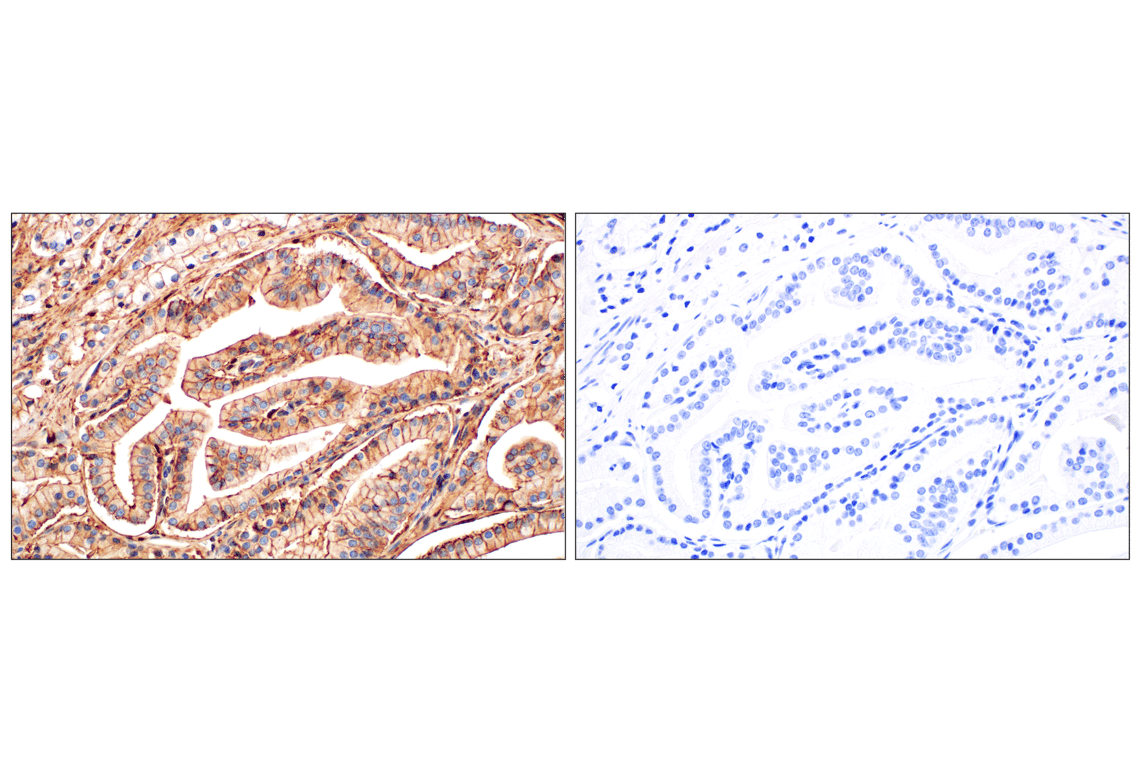
Immunohistochemical analysis of paraffin-embedded human prostate adenocarcinoma using Annexin V (E3W8V) Rabbit mAb (left) compared to concentration-matched Rabbit (DA1E) mAb IgG XP® Isotype Control #3900 (right).
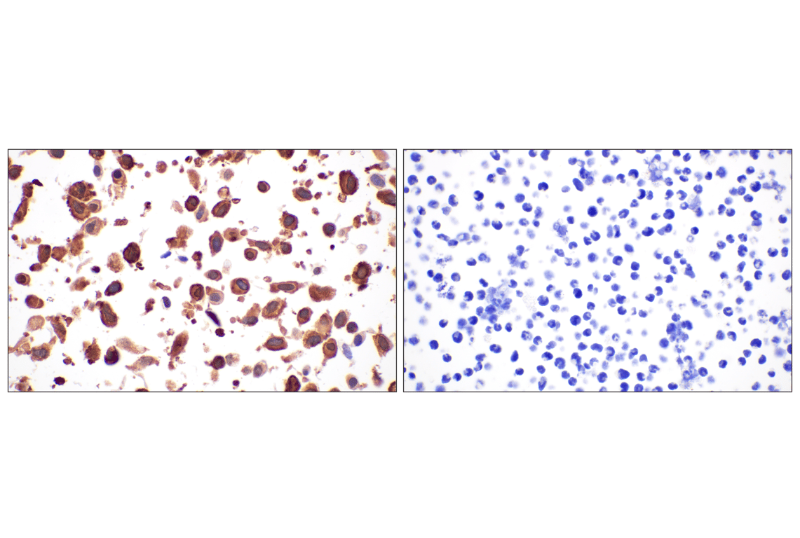
Immunohistochemical analysis of paraffin-embedded TALL-1 cell pellet (left, positive) or U-118 MG cell pellet (right, negative) using Annexin V (E3W8V) Rabbit mAb.
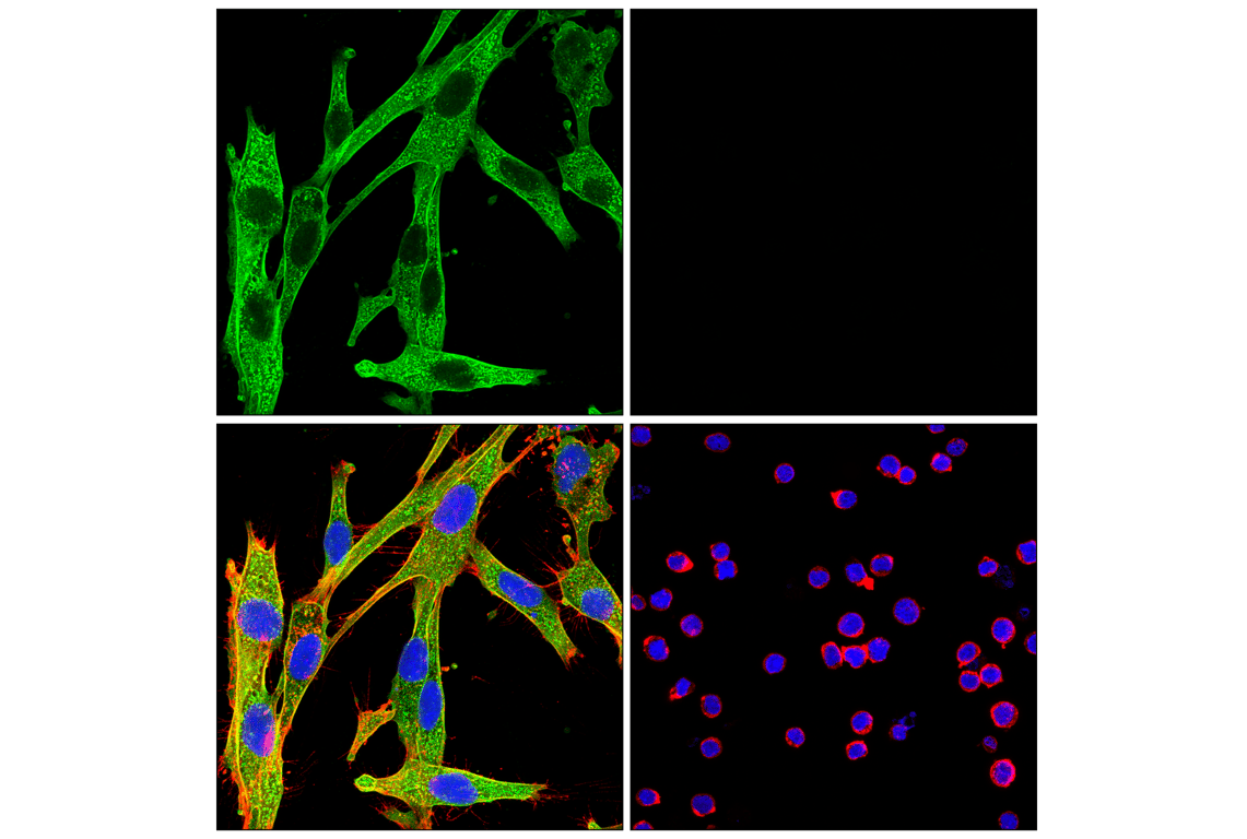
Confocal immunofluorescent analysis of U-118 MG cells (left, positive) and Loucy cells (right, negative) using Annexin V (E3W8V) Rabbit mAb (green), β-Actin (8H10D10) Mouse mAb #3700 (red), and DAPI #4083 (blue).
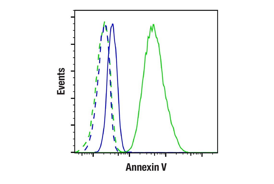
Flow cytometric analysis of TALL-1 cells (blue, negative) and U-118 MG cells (green, positive) using Annexin V (E3W8V) Rabbit mAb (solid lines) or concentration-matched Rabbit (DA1E) mAb IgG XP® Isotype Control #3900 (dashed lines). Anti-rabbit IgG (H+L), F(ab')2 Fragment (Alexa Fluor® 488 Conjugate) #4412 was used as a secondary antibody.