


 全部商品分类


ANTI-HI CYTOKERATIN 7 LP5K PUR

 下载产品说明书 下载COA 下载SDS
 用小程序,查商品更便捷


 收藏
收藏
 对比
对比 咨询
咨询种属反应
已发表种属
宿主/亚型
分类
类型
克隆号
偶联物
形式
浓度
纯化类型
保存液
内含物
保存条件
运输条件
RRID
产品详细信息
Description: This LP5K monoclonal antibody reacts with human cytokeratin 7 (K7), a 54-kDa type II (or basic) keratin expressed either alone or paired with cytokeratin 19 in simple epithelia, mesothelium, urothelium, and pseudostratified epithelium. Expression of cytokeratin 7 in the gastric foveolar, intestinal, and stratified squamous epithelia is extremely low or undetectable. Cytokeratins form the intracellular cytoskeletal network that maintains the integrity and stability of cells and tissues. In addition, most carcinomas express cytokeratin 7. The coordinated expression of this keratin with cytokeratin 20 is commonly used as a diagnostic marker for a variety of carcinomas.
Applications Reported: This LP5K antibody has been reported for use in immunoblotting (WB) and immunocytochemistry (ICC).
Applications Tested: This LP5K antibody has been tested by immunofluorescent staining of paraformaldehyde fixed and permeabilized cells. This can be used at less than or equal to 10 µg/mL. It is recommended that the antibody be titrated for optimal performance in the assay of interest.
Purity: Greater than 90%, as determined by SDS-PAGE.
Aggregation: Less than 10%, as determined by HPLC.
Filtration: 0.2 µm post-manufacturing filtered.
靶标信息
Cytokeratin 7 blocks interferon-dependent interphase and stimulates DNA synthesis in cells. Involved in the translational regulation of the human papillomavirus type 16 E7 mRNA (HPV16 E7).
仅用于科研。不用于诊断过程。未经明确授权不得转售。
生物信息学
蛋白别名: CK-7; cytokeratin 7; Cytokeratin-7; K7; keratin 7, type II; keratin, 55K type II cytoskeletal; keratin, simple epithelial type I, K7; Keratin, type II cytoskeletal 7; Keratin-7; Sarcolectin; type II mesothelial keratin K7; Type-II keratin Kb7
基因别名: CK7; K2C7; K7; KRT7; SCL
UniProt ID:(Human) P08729
Entrez Gene ID:(Human) 3855
参考图片
Immunofluorescence analysis of Cytokeratin 7 was performed using HeLa and SK-OV-3 cells. The cells were fixed with 4% paraformaldehyde for 10 minutes, permeabilized with 0.1% Triton™ X-100 for 15 minutes, and blocked with 2% BSA for 1 hour at room temperature. The cells were labeled with Cytokeratin 7 Monoclonal Antibody (LP5K), eBioscience™ (Product # 14-9005-82) at 5 microgram/mL in 0.1% BSA, incubated at 4 degree celsius overnight and then labeled with Goat anti-Mouse IgG (H+L) Superclonal™ Recombinant Secondary Antibody, Alexa Fluor® 488 (Product # A28175) at a dilution of 1:2000 for 45 minutes at room temperature (Panel a: green). Nuclei (Panel b: blue) were stained with ProLong™ Diamond Antifade Mountant with DAPI (Product # P36962). F-actin (Panel c: red) was stained with Rhodamine Phalloidin (Product # R415, 1:300). Panel d represents the merged image showing expression of Cytokeratin 7 in HeLa. Panel e represents SK-OV-3 cells, showing lesser expression of Cytokeratin 7. Panel f represents control HeLa cells with no primary antibody to assess background. The images were captured at 60X magnification.
Immunocytochemistry of fixed and permeabilized HeLa cells using 10 µg/mL of Mouse IgG2b Isotype Control (Product # 14-4732-82) (left) or Anti-Human Cytokeratin 7 Purified (right) followed by Anti-Mouse TRITC. Nuclei are counterstained with DAPI.
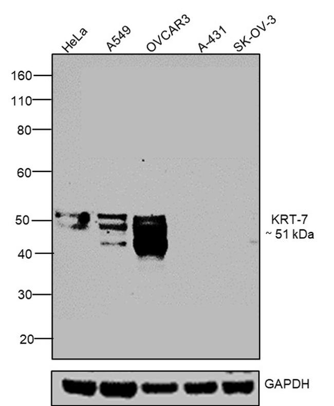
Western blot was performed using Cytokeratin 7 Monoclonal Antibody (LP5K), eBioscience™ (Product # 14-9005-82) and a 51 kDa band corresponding to KRT-7 was observed in positive cell lines such as HeLa, A549, OVCAR-3 and not in negative cell lines such as A431 and SK-OV-3 as mentioned in literature. Membrane enriched extracts (30 µg lysate) of HeLa (Lane 1), A549 (Lane 2), OVCAR-3 (Lane 3), A-431 (Lane 4) and SK-OV-3 (Lane 5) were electrophoresed using NuPAGE® 4-12 % Bis-Tris gel (Product # NP0321BOX). Resolved proteins were then transferred onto a nitrocellulose membrane (Product # IB23001) by iBlot® 2 Dry Blotting System (Product # IB21001). The blot was probed with the primary antibody (1:1000 dilution) and detected by chemiluminescence with Goat anti-Mouse IgG (H+L) Superclonal™ Recombinant Secondary Antibody, HRP (Product # A28177, 1:4000 dilution) using the iBright FL 1000 (Product # A32752). Chemiluminescent detection was performed using Novex® ECL Chemiluminescent Substrate Reagent Kit (Product # WP20005).
Antibody specificity was demonstrated by detection of differential basal expression of the target across tissues owing to their inherent genetic constitution. Higher expression of KRT7 was observed in HeLa, A549, OVCAR-3 cells in comparison to A431 and SK-OV-3 cells using Cytokeratin 7 Monoclonal Antibody (LP5K), eBioscience™ (Product # 14-9005-82) in western blot. {RE}
Antibody specificity was demonstrated by detection of differential basal expression of the target across tissues owing to their inherent genetic constitution. Higher expression of KRT7 was observed in HeLa in comparison to SK-OV-3 cells using Cytokeratin 7 Monoclonal Antibody (LP5K), eBioscience™ (Product # 14-9005-82) in Immunofluorescence. {RE}