


 全部商品分类
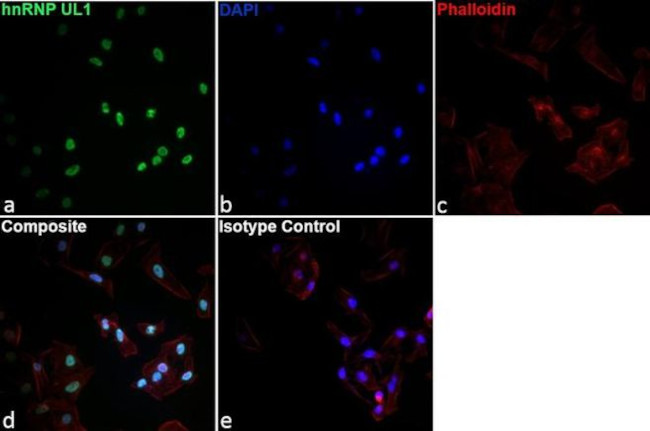


 下载产品说明书 下载COA 下载SDS
 用小程序,查商品更便捷


 收藏
收藏
 对比
对比 咨询
咨询种属反应
宿主/亚型
分类
类型
克隆号
偶联物
形式
浓度
纯化类型
保存液
内含物
保存条件
运输条件
RRID
产品详细信息
Description: This EAP 4A11 monoclonal antibody recognizes human hnRNP UL1. EAP 4A11 detects a ~95 kDa band corresponding to hnRNP UL1 by Western Blot and stains hnRNP UL1 in the nucleus in Immunocytochemistry.
Applications Reported: This EAP 4A11 antibody has been reported for use in western blot and immunocytochemistry/immunofluorescence.
Applications Tested: This EAP 4A11 antibody has been tested by western blot of human cell lysates and immunocytochemistry/immunofluorescence of human cell lines. This may be used at less than or equal to 4.0 µg/ml for western blot and less than or equal to 2.5 µg/mL for immunocytochemistry/immunofluorescence.
靶标信息
HNRPUL1 is a nuclear RNA-binding protein of the heterogeneous nuclear ribonucleoprotein (hnRNP) family. This protein binds specifically to adenovirus E1B-55kDa oncoprotein. It may play an important role in nucleocytoplasmic RNA transport, and its function is modulated by E1B-55kDa in adenovirus-infected cells.This gene encodes a nuclear RNA-binding protein of the heterogeneous nuclear ribonucleoprotein (hnRNP) family. This protein binds specifically to adenovirus E1B-55kDa oncoprotein. It may play an important role in nucleocytoplasmic RNA transport, and its function is modulated by E1B-55kDa in adenovirus-infected cells. Four transcript variants encoding different isoforms have been found for this gene. Another variant has also been found, but its full-length nature has not been determined.
仅用于科研。不用于诊断过程。未经明确授权不得转售。
生物信息学
蛋白别名: Adenovirus early region 1B-associated protein 5; E1B 55kDa associated protein 5; E1B-55 kDa-associated protein 5; E1B-AP5; heterogeneous nuclear ribonucleoprotein U-like 1; Heterogeneous nuclear ribonucleoprotein U-like protein 1
基因别名: E1B-AP5; E1BAP5; HNRNPUL1; HNRPUL1
UniProt ID:(Human) Q9BUJ2
Entrez Gene ID:(Human) 11100
参考图片
Immunofluorescence analysis of hnRNP UL1 Monoclonal Antibody (Product # 14-9806-82) was performed using 70% confluent log phase HeLa cells. The cells were fixed with 4% paraformaldehyde for 15 minutes at room temperature, permeabilized with 0.1% Triton X-100 for 30 minutes at 37°C, and blocked with 10% Normal Goat Serum for 1 hour at room temperature. The cells were labeled with 1.25 µg/mL of hnRNP UL1 Monoclonal Antibody (Product # 14-9806-82) in 0.1% Triton X-100, incubated overnight at 4°C and then labeled with 4 µg/mL of Goat anti-Rat IgG (H+L) Cross-Adsorbed Secondary Antibody, Alexa Fluor 488 conjugate (Product # A11006) for 1 hour at room temperature (Panel a: green). Nuclei (Panel b: blue) were stained with Fluoromount G with DAPI (Product # 00-4959). F-actin (Panel c: red) was stained with Rhodamine Phalloidin (Product # R415, 1:300). Panel d represents the merged image showing nuclear localization. Panel e represents a merged image of control cells incubated with 1.25 µg/mL of Rat IgG2b Isotype Control (Product # 14-4031-85) antibody to assess background. The images were captured at 40X magnification.
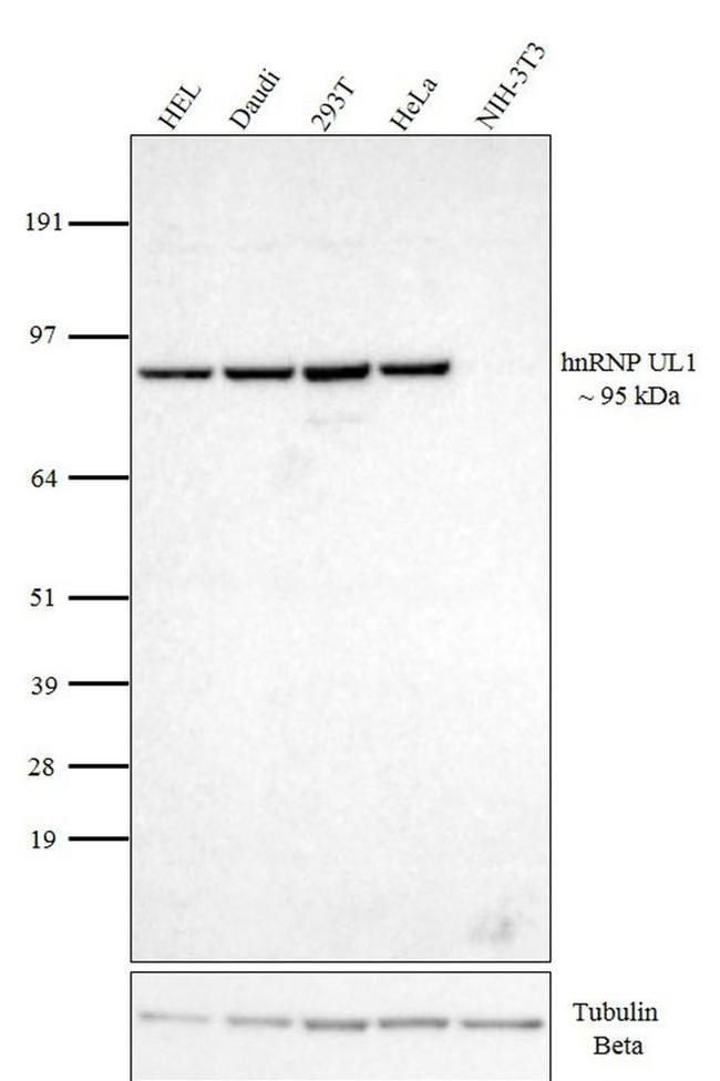
Western Blot analysis was performed on whole cell extracts using 10 µg lysate of HEL (Lane 1), Daudi (Lane 2), 293T (Lane 3), HeLa (Lane 4), and NIH 3T3 (Lane 5). The blot was probed with Anti-hnRNP UL1 Monoclonal antibody clone EAP 4A11 (2 µg/ml) and detected by chemiluminescence using Goat-anti Rat IgG (H+L) Secondary Antibody, HRP (Product # 31471) at a 1: 4,000 dilution. Visualization using SuperSignal™ West Pico PLUS Chemiluminescent Substrate (Product # 34578). The blot was reprobed with beta Tubulin Loading Control Antibody (Product # MA5-16308) and Goat anti-Mouse IgG (H+L) Secondary Antibody, HRP (Product # 31430)