


 全部商品分类


ANTI-HU IGG FCG-SP PE

 下载产品说明书 下载COA 下载SDS
 用小程序,查商品更便捷


 收藏
收藏
 对比
对比 咨询
咨询
流式细胞分析 (Flow)
种属反应
宿主/亚型
分类
类型
偶联物
激发/发射光谱

形式
浓度
纯化类型
保存液
内含物
保存条件
运输条件
RRID
靶标
抗体形式
产品详细信息
Description: This goat antibody reacts with the Fc fragment of heavy chains on human IgG but notwith the light chains on most human immunoglobulins. No antibody was detected against human IgM or IgA, or against nonimmunoglobulinserum proteins. This antibody has been tested to ensure minimalcross-reaction with bovine, horse, and mouse serum proteins, but the antibody may cross-react with immunoglobulins fromother species.
Applications Reported: PE goat anti-Human IgG (Fc gamma Fragment Specific) has been reported for use in flow cytometric analysis.
Applications Tested: PE goat anti-Human IgG (Fc gamma Fragment Specific) has been tested by flow cytometric analysis to detect purified human Fc gamma-tagged recombinant proteins. This can be used at less than or equal to 1 µg per test. A test is defined as the amount (µg) of antibody that will stain a cell sample in a final volume of 100 µL. Cell number should be determined empirically but can range from 10^5 to 10^8 cells/test. It is recommended that the antibody be carefully titrated for optimal performance in the assay of interest.
Excitation: 488-561 nm; Emission: 578 nm; Laser: Blue Laser, Green Laser, Yellow-Green Laser.
Filtration: 0.2 µm post-manufacturing filtered.
靶标信息
Anti-Human secondary antibodies are affinity-purified antibodies with well-characterized specificity for human immunoglobulins and are useful in the detection, sorting or purification of its specified target. Secondary antibodies offer increased versatility enabling users to use many detection systems (e.g. HRP, AP, fluorescence). They can also provide greater sensitivity through signal amplification as multiple secondary antibodies can bind to a single primary antibody. Most commonly, secondary antibodies are generated by immunizing the host animal with a pooled population of immunoglobulins from the target species and can be further purified and modified (i.e. immunoaffinity chromatography, antibody fragmentation, label conjugation, etc.) to generate highly specific reagents.
仅用于科研。不用于诊断过程。未经明确授权不得转售。
参考图片
C57BL/6 mouse splenocytes were incubated with Mouse CCL19 (MIP-3 beta) Recombinant Protein (a human IgG Fc-tagged ligand that interacts with CCR7), followed by CD4 Monoclonal Antibody, APC (Product # 17-0041-82) and staining buffer (left) or 0.5 µg of Anti-IgG Fc Secondary Antibody, PE (right). Cells in the lymphocyte gate were used for analysis.
Anti-Siglec-15 blocking mAb 5G12 competes for the sialic acid-binding site of Siglec-15.a Representative flow cytometry histograms of activated CD8+ and CD4+ human T cells showing the binding of recombinant Siglec-15-Fc in the presence or absence of anti-Siglec-15 5G12 Fab. Here, secondary control means that only an anti-Fc detector antibody, but not recombinant Fc-chimera protein was added to the sample. b Bar graphs show Siglec-15-Fc binding to human T cells in the presence or absence of 5G12 Fab (CD4+: 0.0001, CD8+: 0.0002, n = 4 donors). Errors bars denote SEM. ***p < 0.001 as determined by two-tailed, unpaired Student’s t test. c Competition of 3′SL (left)/6′SL (right) and 5G12 mAb for the same binding site of Siglec-15. Source data are provided as a Source Data file. - Image collected and cropped by CiteAb under a CC-BY license from the following publication: Structural insights into Siglec-15 reveal glycosylation dependency for its interaction with T cells through integrin CD11b.
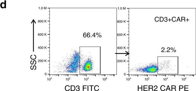
Clinical and pathological findings after autologous HER2 CAR T-cell infusions.a Histological examination of the bone marrow 4 weeks after the salvage chemotherapy (ARST0921) and prior to initiating CAR T-cell infusions showing hypocellularity and presence of rhabdomyosarcoma (RMS) cells on hematoxylin and eosin (H&E) staining and immunoreactivity to desmin and myogenin, b complete disease response (CR1), evidenced by recovery of trilineage hematopoiesis and absence of immunoreactivity to desmin and myogenin after three HER2 CAR T-cell infusions. Panels (a, b) show representative microscopic images; scale bar 100 µm. c Representative image from positron emission tomography-computed tomography (PET-CT) with no evidence of FDG-avid disease in bone marrow or other sites 6 weeks after the third HER2 CAR T-cell infusion. d Detection of HER2 CAR-expressing T cells in the peripheral blood 7 days after the second infusion using flow cytometry. HER2 CAR was specifically recognized using HER2.Fc chimeric protein followed by a goat anti-human Fc conjugated with PE as a secondary antibody. SSC side scatter. e The proportion of CD3+ HER2 CAR-expressing T cells on day +7 after each infusion during the induction period. f Histograms showing the PD-1 and LAG3 surface expression in CAR-positive CD8+ (in blue) in comparison to CAR-negative CD8+ T cells (in black) at peak expansion (day +7) after each infusion during induction, and g the… Image collected and cropped by CiteAb from the following publication (https://pubmed.ncbi.nlm.nih.gov/32669548), licensed under a CC BY license.
Validation of ADCs. (A) hROR2 surface expression in cell lines used in this study as determined by FACS using the hROR2-specific antibody XBR2-401 (37) detected using a PE-labeled anti-human Fcγ. Black histograms, ROR2 staining; gray histograms, control staining with only the secondary antibody. (B) Following recovery of VH and VL sequences by RT-PCR cloning from hROR2-ECD-Twin-Strep reactive Transpo-mAb cell clones and expression in HEK293T cells, purified recombinant antibodies were site-specifically conjugated to a derivative of the potent cytotoxic payload PNU-159682 by sortase-mediated antibody conjugation (SMAC-technologyTM) to the C-termini of the heavy and light chain constant domains. These ADCs were tested for their cell killing activity on hROR2 negative (L363), and hROR2-high (T47D-hROR2) cell lines. Viability of the cells is plotted in arbitrary units (a.u.) of luminescence on the y-axis as a function of the concentration of the ADCs on the x-axis as a result of a CellTiter-Glo cell-killing assay. hROR2-negative L363 cells were used as negative controls (with an isotype-control mAb in light gray that binds to a TAA on L363). G3 and G5 designate the length of a glycine linker used in the ADCs, which has previously been shown not to affect ADC potency. Datapoints represent mean of two replicates and error bars represent SD. Image collected and cropped by CiteAb from the following publication (https://pubmed.ncbi.nlm.nih.gov/30450096), licensed under a CC BY license.