


 全部商品分类


ANTI-MO GRANZYME B 16G6 PUR

 下载产品说明书 下载COA 下载SDS
 用小程序,查商品更便捷


 收藏
收藏
 对比
对比 咨询
咨询
种属反应
已发表种属
宿主/亚型
分类
类型
克隆号
偶联物
形式
浓度
纯化类型
保存液
内含物
保存条件
运输条件
RRID
产品详细信息
Description: The 16G6 antibody reacts with mouse Granzyme B (GrB) which is a member of the granzyme serine protease family. GrB is found in the granules of cytotoxic T cells and NK cells. Granzyme B has also been described as CGL1 (cathepsin G-like-1), a serine protease expressed only in cytotoxic T-lymphocytes after cell activation. GrB has been called CTLA-1 (cytotoxic T lymphocyte-associated serine esterase 1) based on identification of mRNA in various cytotoxic T cells, but not observed in non-cytotoxic lymphoid cells. GrB is crucial for the rapid induction of target cell death by apoptosis, induced by interaction with cytotoxic T cells. The receptor involved has been identified as mannose 6-phosphate receptor. This receptor functions as a death receptor for granzyme B during cytotoxic T cell-induced apoptosis.
For intracellular staining and flow cytometric analysis with direct conjugates of anti-mouse Granzyme B, it is highly recommended to use the Foxp3 Staining Buffer Set (Product # 00-5523). Other buffers may yield varying results.
Applications Reported: This 16G6 antibody has been reported for use in western blotting, immunohistochemical staining, ELISA capture antibody, and immunohistochemical staining of formalin-fixed paraffin embedded tissue sections.
Applications Tested: The unlabelled 16G6 antibody has been tested as the capture antibody in a sandwich ELISA for quantitation of mouse granzyme B protein levels, in combination with the biotinylated eBioLUEE (Product # 13-8821-81) antibody for detection and recombinant mouse granzyme B as the standard. A suitable range of concentrations of this antibody for ELISA detection is 1-4 µg/mL.
The 16G6 antibody has been tested for Western blotting; a recommended starting concentration is 1 µg/mL. The 16G6 antibody has been used for formalin-fixed paraffin embedded tissue using low pH antigen retrieval at less than or equal to 20 µg/mL. It is recommended that the antibody be carefully titrated for optimal performance in the assay of interest.
Purity: Greater than 90%, as determined by SDS-PAGE.
Aggregation: Less than 10%, as determined by HPLC.
Filtration: 0.2 µm post-manufacturing filtered.
靶标信息
Granzyme B is a member of the granzyme serine protease family, and is found in the granules of cytotoxic T cells and NK cells. Granzyme B has been described as CGL1 (cathepsin G-like-1), a serine protease expressed only in cytotoxic T-lymphocytes after cell activation, and CTLA-1 (cytotoxic T lymphocyte-associated serine esterase 1) based on identification of mRNA in various cytotoxic T cells, but not observed in non-cytotoxic lymphoid cells. Granzyme B is crucial for the rapid induction of target cell death by apoptosis, induced by interaction with cytotoxic T cells. The receptor involved in this process has been identified as mannose 6-phosphate receptor which functions as a death receptor for Granzyme B during cytotoxic T cell-induced apoptosis. Granzyme B enters target cells to cleave caspase-3 and initiate the caspase cascade leading to DNA fragmentation and apoptosis. Granzyme B can also act through a mitochondrial apoptosis pathway by cleaving the Bid protein. Granzymes are neutral serine proteases, which are stored in specialized lytic granules of cytotoxic T lymphocytes (CTLs) and in natural killer (NK) cells. A number of granzymes (A to G) have been isolated and cloned from mouse CTLs and NK cells, however in man, fewer have been cloned and identified.
仅用于科研。不用于诊断过程。未经明确授权不得转售。
生物信息学
蛋白别名: C11; Cathepsin G-like 1; CCP1; CTLA-1; CTSGL1; Cytotoxic cell protease 1; cytotoxic serine protease B; Cytotoxic T lymphocyte associated serine esterase 1; Cytotoxic T-lymphocyte proteinase 2; cytotoxic T-lymphocyte-associated serine esterase 1; fragmentin 2; Fragmentin-2; granzyme 2; Granzyme B; granzyme B (granzyme 2, cytotoxic T-lymphocyte-associated serine esterase 1); Granzyme B(G,H); Granzyme-2; GranzymeB; HLP; Human lymphocyte protein; Human lymphocyte protein (Hlp); Lymphocyte protease; OTTHUMP00000028189; SECT; T-cell serine protease 1-3E
基因别名: AI553453; CCP-1/C11; CCP1; CCPI; CGL-1; CGL1; CSP-B; CSPB; Ctla-1; CTLA1; CTSGL1; GRB; GZB; GZMB; HLP; SECT
UniProt ID:(Human) P10144, (Mouse) P04187
Entrez Gene ID:(Human) 3002, (Mouse) 14939
参考图片
Immunohistochemical analysis of Granzyme B was performed using formalin-fixed paraffin-embedded human tonsil tissue sections. To expose the target protein, heat-induced epitope retrieval was performed on de-paraffinized sections using eBioscience™ IHC Antigen Retrieval Solution - High pH (10X) (Product # 00-4956-58) diluted to 1X solution in water in a decloaking chamber at 110 degree Celsius for 15 minutes. Following antigen retrieval, the sections were blocked with 3% H2O2 for 1h at room temperature followed by 2% normal goat serum in 1X PBS for 45 minutes at room temperature and then probed with or without Granzyme B Monoclonal Antibody (16G6), eBioscience™ (Product # 14-8822-82) at 1 µg/mL in 0.1% normal goat serum overnight at 4 degree Celsius in a humidified chamber. Detection was performed using F(ab)2-Rabbit anti-Rat IgG (H+L) Secondary Antibody, HRP (Product # PA1-29927) (1:1500 dilution) and Alexa Fluor™ 594 Tyramide conjugate (Product # B40957). Nuclei were stained with DAPI (Product # D1306) and the sections were mounted using ProLong™ Glass Antifade Mountant (Product # P36984). The images were captured on EVOS™ M7000 Imaging System (Product # AMF7000) at 20X magnification and externally deconvoluted.
Immunohistochemical analysis of Granzyme B was performed using formalin-fixed paraffin-embedded human tonsil tissue sections. To expose the target protein, heat-induced epitope retrieval was performed on de-paraffinized sections using eBioscience™ IHC Antigen Retrieval Solution - High pH (10X) (Product # 00-4956-58) diluted to 1X solution in water in a decloaking chamber at 110 degree Celsius for 15 minutes. Following antigen retrieval, the sections were blocked with 3% H2O2 for 1h at room temperature followed by 2% normal goat serum in 1X PBS for 45 minutes at room temperature and then probed with or without Granzyme B Monoclonal Antibody (16G6), eBioscience™ (Product # 14-8822-82) at 1 µg/mL in 0.1% normal goat serum overnight at 4 degree Celsius in a humidified chamber. Detection was performed using F(ab)2-Rabbit anti-Rat IgG (H+L) Secondary Antibody, HRP (Product # PA1-29927) (1:1500 dilution) and Alexa Fluor™ 594 Tyramide conjugate (Product # B40957). Nuclei were stained with DAPI (Product # D1306) and the sections were mounted using ProLong™ Glass Antifade Mountant (Product # P36984). The images were captured on EVOS™ M7000 Imaging System (Product # AMF7000) at 20X magnification and externally deconvoluted.
Immunohistochemical analysis of Granzyme B was performed using formalin-fixed paraffin-embedded human tonsil tissue sections. To expose the target protein, heat-induced epitope retrieval was performed on de-paraffinized sections using eBioscience™ IHC Antigen Retrieval Solution - Low pH (10X) (Product # 00-4955-58) diluted to 1X solution in water in a decloaking chamber at 110 degree Celsius for 15 minutes. Following antigen retrieval, the sections were blocked with 3% H2O2 for 1h at room temperature followed by 2% in 1X PBS for 45 minutes at room temperature and then probed with or without Granzyme B Monoclonal Antibody (16G6), eBioscience™ (Product # 14-8822-82) at 1 µg/mL in 0.1% overnight at 4 degree Celsius in a humidified chamber. Detection was performed using F(ab)2-Rabbit anti-Rat IgG (H+L) Secondary Antibody, HRP (Product # PA1-29927) (1:1500 dilution) and Alexa Fluor™ 594 Tyramide conjugate (Product # B40957). Nuclei were stained with DAPI (Product # D1306) and the sections were mounted using ProLong™ Glass Antifade Mountant (Product # P36984). The images were captured on EVOS™ M7000 Imaging System (Product # AMF7000) at 20X magnification and externally deconvoluted.
Immunohistochemical analysis of Granzyme B was performed using formalin-fixed paraffin-embedded human tonsil tissue sections. To expose the target protein, heat-induced epitope retrieval was performed on de-paraffinized sections using eBioscience™ IHC Antigen Retrieval Solution - Low pH (10X) (Product # 00-4955-58) diluted to 1X solution in water in a decloaking chamber at 110 degree Celsius for 15 minutes. Following antigen retrieval, the sections were blocked with 3% H2O2 for 1h at room temperature followed by 2% in 1X PBS for 45 minutes at room temperature and then probed with or without Granzyme B Monoclonal Antibody (16G6), eBioscience™ (Product # 14-8822-82) at 1 µg/mL in 0.1% overnight at 4 degree Celsius in a humidified chamber. Detection was performed using F(ab)2-Rabbit anti-Rat IgG (H+L) Secondary Antibody, HRP (Product # PA1-29927) (1:1500 dilution) and Alexa Fluor™ 594 Tyramide conjugate (Product # B40957). Nuclei were stained with DAPI (Product # D1306) and the sections were mounted using ProLong™ Glass Antifade Mountant (Product # P36984). The images were captured on EVOS™ M7000 Imaging System (Product # AMF7000) at 20X magnification and externally deconvoluted.
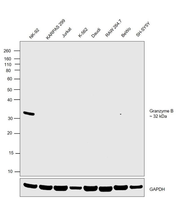
Western blot was performed using Anti-Granzyme B Monoclonal Antibody (16G6), eBioscience™ (Product # 14-8822-80, 14-8822-82) and a 32 kDa band corresponding to Granzyme B was observed only in NK-92 cells which is reported to be positive and not in any other cell lines. Whole cell extracts (30 µg lysate) of NK-92 (Lane 1), KARPAS 299 (Lane 2), Jurkat (Lane 3), K-562 (Lane 4), Daudi (Lane 5), RAW 264.7 (Lane 6), BeWo (Lane 7) and SH-SY5Y (Lane 8) were electrophoresed using NuPAGE™ 10% Bis-Tris Protein Gel (Product # NP0302BOX). Resolved proteins were then transferred onto a nitrocellulose membrane (Product # IB23001) by iBlot® 2 Dry Blotting System (Product # IB21001). The blot was probed with the primary antibody (1 µg/mL) and detected by chemiluminescence with F(ab"e2)-Rabbit anti-Rat IgG (H+L) Secondary Antibody, HRP (Product # PA1-29927,1:4000 dilution) using the iBright FL 1000 (Product # A32752). Chemiluminescent detection was performed using Novex® ECL Chemiluminescent Substrate Reagent Kit (Product # WP20005).
BALB/c splenocytes were stimulated with Mouse IL-2 Recombinant (100 ng/mL) (Product # 14-8021-64) for 3 days. Subsequently, splenocyte lysates were loaded at 1x10e6 cells/lane, probed with 1 µg/mL Anti-Mouse Granzyme B Purified and revealed with Anti-Rat IgG HRP.
Figure 4 SFIH combined with an anti-PD-1 antibody alters the immune landscape in the tumor microenvironment. Immunohistochemistry (IHC) staining of MC38 tumors for infiltration of immune cells. CD3, CD8, granzyme B, F4/80, and TGF-beta1 levels were assessed by microscopic examination of IHC-stained sections. Representative IHC-stained images are shown (scale bars = 100 um).
Figure 3 CD73 presents a negative spatial correlation with anti-tumor immunity. (A, C) Analysis of Granzyme B IHC staining from RFA + AB680 treated tumors 10 days after ablation revealed increased abundance of Granzyme B + cells when compared to KPC control tumors subcutaneously grown up to 10 days (p < 0.0001), RFA (p < 0.01) and RFA+VEH (p < 0.01) treated mice . (B, D) Analysis of CD8alpha positive cells IHC staining from RFA + AB680 treated tumors 10 days after ablation revealed increased abundance of CD8alpha + cells when compared to KPC control tumors subcutaneously grown up to 10 days, RFA and RFA+VEH treated mice (p < 0.0001). (E) At 10 days after treatment, a negative correlation was observed between CD73 protein expression (Left) and abundance of Granzyme B + cells (Right) in composite pictures of RFA treated KPC subcutaneous tumors taken at 4X after IHC. (F) Higher magnification images reveal areas with decreased CD73 expression present increased granzyme B+ cells (Left panels, a and b), whereas areas with increased CD73 expression show decreased Granzyme B+ cells (Right panels, c and d), suggesting elevated antitumor immunity in areas with lower ADO generation. One-way ANOVA (C, D) were used for group comparisons. Bars represent 50uM. **p<0.01; ****p<0.0001; ns (not significant).
Published figure using Granzyme B monoclonal antibody (Product # 14-8822-82) in Immunohistochemistry
Published figure using Granzyme B monoclonal antibody (Product # 14-8822-82) in Western Blot
Published figure using Granzyme B monoclonal antibody (Product # 14-8822-82) in Western Blot
Published figure using Granzyme B monoclonal antibody (Product # 14-8822-82) in Western Blot
Published figure using Granzyme B monoclonal antibody (Product # 14-8822-82) in Flow Cytometry
Antibody specificity was demonstrated by detection of differential basal expression of the target across cell lines owing to their inherent genetic constitution. Relative expression of Granzyme B was observed only in NK-92 cells which is reported to be positive and not in any other cell lines using Anti-Granzyme B Monoclonal Antibody (16G6), eBioscience™ (Product # 14-8822-80, 14-8822-82) in Western Blot. {RE}