


 全部商品分类
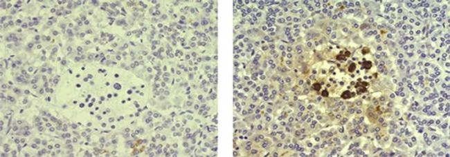


ANTI-SOMATOSTATIN ICDCLS PUR

 下载产品说明书 下载COA 下载SDS
 用小程序,查商品更便捷


 收藏
收藏
 对比
对比 咨询
咨询种属反应
已发表种属
宿主/亚型
分类
类型
克隆号
偶联物
形式
浓度
纯化类型
保存液
内含物
保存条件
运输条件
RRID
产品详细信息
Description: The monoclonal antibody ICDCLS recognizes human, mouse, and rat Somatostatin. Somatostatin, also known as growth hormone-inhibiting hormone (GHIH) or somatotrophin release-inhibiting factor (SRIF), is a peptide hormone with two active forms (14 and 28 amino acids) produced by alternative cleavage of a single preproprotein. Somatostatin is secreted by the delta cells of the islet of Langerhans in the pancreas as well as by the hypothalamus and intestine. Somatostatin has a variety of functions, such as regulating the endocrine system and affecting neurotransmission and cell proliferation. Somatostatin also inhibits the release of numerous secondary hormones, such as insulin and glucagon. Somatostatin can be used to identify medium size aspiny neurons in the striatum, cortex, and hippocampus. Expression of somatostatin is present in hyperplasia of the pancreatic islets as well as islet cell tumors such as somatostatinomas.
Applications Reported: This ICDCLS antibody has been reported for use in immunohistochemical staining of formalin-fixed paraffin embedded tissue sections, and microscopy.
Applications Tested: This ICDCLS antibody has been tested by immunohistochemistry of formalin-fixed paraffin embedded human tissue using low pH antigen retrieval and can be used at less than or equal to 0.1 µg/mL. It is recommended that the antibody be carefully titrated for optimal performance in the assay of interest.
Purity: Greater than 90%, as determined by SDS-PAGE.
Aggregation: Less than 10%, as determined by HPLC.
Filtration: 0.2 µm post-manufacturing filtered.
靶标信息
The hormone somatostatin has active 14 aa and 28 aa forms that are produced by alternate cleavage of the single preproprotein encoded by this gene. Somatostatin is expressed throughout the body and inhibits the release of numerous secondary hormones by binding to high-affinity G-protein-coupled somatostatin receptors. This hormone is an important regulator of the endocrine system through its interactions with pituitary growth hormone, thyroid stimulating hormone, and most hormones of the gastrointestinal tract. Somatostatin also affects rates of neurotransmission in the central nervous system and proliferation of both normal and tumorigenic cells.
仅用于科研。不用于诊断过程。未经明确授权不得转售。
生物信息学
蛋白别名: Growth hormone release-inhibiting factor; prepro-somatostatin; preprosomatostatin; Somatostatin; somatostatin 28; somatostatin-14; somatostatin-28; somatotropin release-inhibiting factor; SRIF
基因别名: SMST; SOM; SRIF; SS; SS-14; SS-28; SST
UniProt ID:(Human) P61278, (Mouse) P60041, (Rat) P60042
Entrez Gene ID:(Human) 6750, (Mouse) 20604, (Rat) 24797
参考图片
Immunohistochemistry of formalin-fixed paraffin embedded human pancreas tissue using 0.1 µg/mL of Mouse IgG1 K Isotype Control Purified (left) or 0.1 µg/mL of Anti-Somatostatin Purified (right) followed by Anti-Mouse IgG Biotin, Streptavidin-HRP, and DAB visualization.Nuclei are counterstained with hematoxylin.
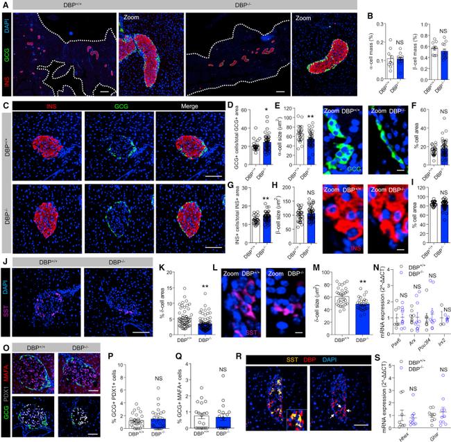
Figure 2 DBP Alters alpha Cell and delta Cell Number and Size (A and B) Cell resolution reconstruction of pancreatic sections reveals no differences in alpha cell and beta cell mass in DBP +/+ and DBP -/- mice (A), quantified in bar graph (B) (scale bar represents 530 mum; representative images are shown; inset is a zoom showing maintenance of cellular resolution in a single image; n = 9-12 sections from 3 to 4 animals; unpaired t test). (C-I) Morphological analyses of DBP -/- islets (C) reveal increased alpha cell number (D) and decreased alpha cell size (E) (representative images shown in right panel), but normal area occupied (F). By contrast, beta cell number is increased (G), although beta cell size (H) (representative images shown in right panel) and area (I) are unchanged (scale bar in D represents 85 mum; scale bars in E and H represent 10 mum; n = 24-45 islets from 3 to 4 animals; E and H are zooms of C to better show alpha cell and beta cell size; DAPI is shown in blue; unpaired t test). (J-M) delta cell proportion (J and K; n = 55-79 islets from 4 to 5 animals) and size (L and M; n = 29 to 30 islets from 3 animals) are decreased in DBP -/- islets (scale bar in J represents 85 mum; scale bar in L represents 10 mum; representative images are shown; L is a zoom of J to better show delta cell size; unpaired t test). (N) Expression levels of the alpha cell differentiation markers Arx , Pax6 , Pou3f4 , and Irx2 are similar in DBP +/- and DBP -/- islets (n = 3-10 animals;
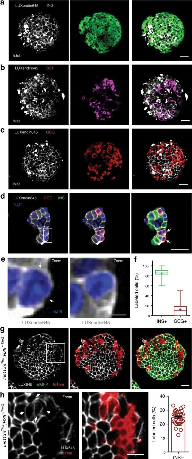
Fig. 4 LUXendin645 reveals GLP1R expression in a subpopulation of alpha-cells. a - c LUXendin645 labeling is widespread throughout the intact islet, co-localizing predominantly with beta-cells a and delta-cells b , but less so with alpha-cells c stained for insulin (INS), somatostatin (SST), and glucagon (GCG), respectively ( n = 18 islets, seven animals, three separate islet preparations) (scale bar = 26 um). d Following dissociation of islets into cell clusters, LUXendin645 labeling can be more accurately quantified (arrows highlight cells selected for zoom-in) (scale bar = 26 um). e Zoom-in of d showing a LUXendin645 - (left) and LUXendin645 + (right) alpha-cell (arrows highlight non-labeled cell membrane, which is not bounded by a beta-cell) (scale bar = 26 um). f Box-and-whiskers plot showing proportion of beta-cells (INS) and alpha-cells (GCG) co-localized with LUXendin645 ( n = 18 cell clusters, ten animals, three separate islet preparations) (box and whiskers plot shows range and median; mean is shown by a plus symbol). g Ins1Cre Thor ;R26 mT/mG dual fluorophore reporter islets express tdTomato until Cre-mediated replacement with mGFP, allowing identification of beta-cells (~80% of the islet population) and non-beta-cells for live imaging (scale bar = 26 um). LUXendin645 (LUX645) highlights GLP1R expression in nearly all beta-cells but relatively few non-beta-cells ( n = 31 islets, six animals, three separate islet preparations). h A zoom