


 全部商品分类


 用小程序,查商品更便捷


Monoclonal antibody is produced by immunizing animals with recombinant mouse ASC/TMS1 protein.
Product Usage Information
| Application | Dilution |
|---|---|
| Western Blotting | 1:1000 |
| Immunoprecipitation | 1:100 |
| Immunohistochemistry (Paraffin) | 1:100 - 1:400 |
| Immunofluorescence (Frozen) | 1:400 - 1:1600 |
| Immunofluorescence (Immunocytochemistry) | 1:400 - 1:1600 |
| Flow Cytometry (Fixed/Permeabilized) | 1:400 - 1:1600 |
Specificity/Sensitivity
Species Reactivity:
Mouse
Supplied in 10 mM sodium HEPES (pH 7.5), 150 mM NaCl, 100 µg/ml BSA, 50% glycerol and less than 0.02% sodium azide. Store at –20°C. Do not aliquot the antibody.
For a carrier free (BSA and azide free) version of this product see product #37953.
参考图片
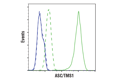
Flow cytometric analysis of Raw264.7 cells (blue) and J774A.1 cells (green) using ASC/TMS1 (D2W8U) Rabbit mAb (solid lines) or a concentration-matched Rabbit (DA1E) mAb IgG XP® Isotype Control #3900 (dashed lines). Anti-rabbit IgG (H+L), F(ab')2 Fragment (Alexa Fluor® 488 Conjugate) #4412 was used as a secondary antibody.
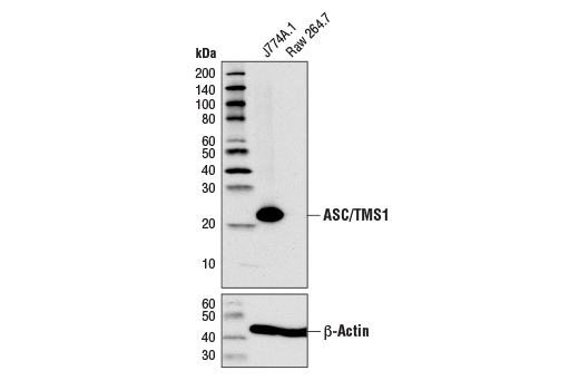
Western blot analysis of extracts from J774A.1 and Raw 264.7 cells using ASC/TMS1 (D2W8U) Rabbit mAb (upper) or β-Actin (D6A8) Rabbit mAb #8457 (lower).
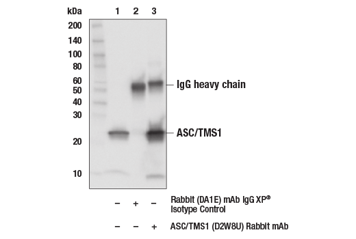
Immunoprecipitation of ASC/TMS1 from J774A.1 cell extracts. Lane 1 is 10% input, lane 2 is Rabbit (DA1E) mAb IgG XP® Isotype Control #3900, and lane 3 is ASC (D2W8U) Rabbit mAb. Western blot analysis was performed using ASC/TMS1 (D2W8U) Rabbit mAb.
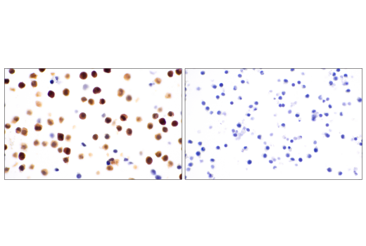
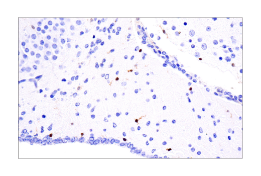
Immunohistochemical analysis of paraffin-embedded J774A.1 cell pellet (left, positive) or RAW 264.7 cell pellet (right, negative) using ASC/TMS1 (D2W8U) Rabbit mAb.
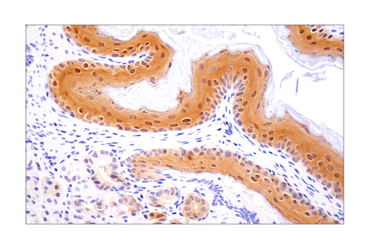
Immunohistochemical analysis of paraffin-embedded mouse forestomach using ASC/TMS1 (D2W8U) Rabbit mAb.
Immunohistochemical analysis of paraffin-embedded mouse brain using ASC/TMS1 (D2W8U) Rabbit mAb.
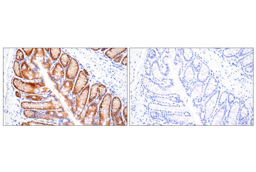
Immunohistochemical analysis of paraffin-embedded mouse colon using ASC/TMS1 (D2W8U) Rabbit mAb (left) compared to concentration-matched Rabbit (DA1E) mAb IgG XP® Isotype Control #3900 (right).
Immunohistochemical analysis of paraffin-embedded mouse thymus using ASC/TMS1 (D2W8U) Rabbit mAb.
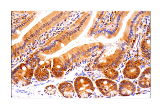
Immunohistochemical analysis of paraffin-embedded mouse small intestine using ASC/TMS1 (D2W8U) Rabbit mAb.
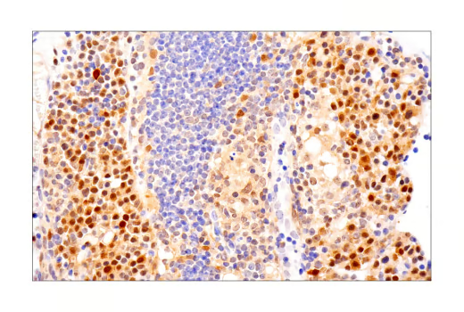
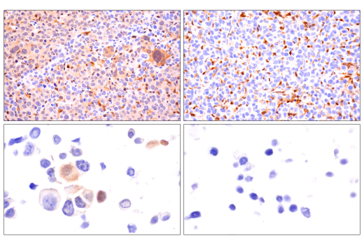
Immunohistochemical analysis of paraffin-embedded Renca syngeneic tumor (top left), 4T1 syngeneic mammary tumor (top right), Renca cell pellet (bottom left), and 4T1 cell pellet (bottom right) using ASC/TMS1 (D2W8U) Rabbit mAb. Both tumors show staining of infiltrating immune cells. Note the presence of staining in the Renca tumor cells and the lack of staining in the 4T1 tumor cells consistent with staining results on corresponding cell pellets.
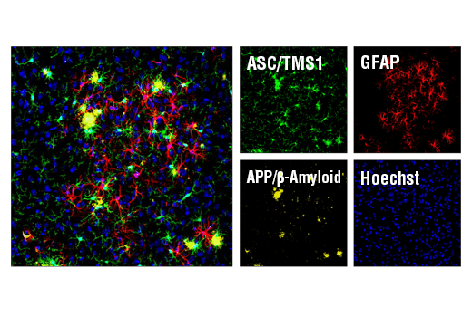
Confocal immunofluorescent analysis of mouse Tg2576 brain which overexpresses mutant human APP695. Sections were first labeled with ASC/TMS1 (D2W8U) Rabbit mAb #67824 (green) and APP/β-Amyloid (NAB228) Mouse mAb #2450 (yellow). After blocking free secondary binding sites with Mouse (G3A1) mAb IgG1 Isotype Control #5415, sections were incubated with GFAP (GA5) Mouse mAb (Alexa Fluor® 647 Conjugate) #3657 (red). Nuclei were labeled with Hoechst 33342 #4082 (blue).
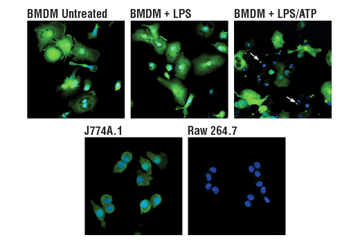
Confocal immunofluorescent analysis of mouse primary bone marrow-derived macrophages (BMDMs) either untreated (upper left) or treated with LPS (50 ng/ml, 4 hr, middle) or LPS followed by ATP (5 mM, 45 min, upper right), and J774A.1 (lower left) or Raw 264.7 (lower right) cells, using ASC/TMS1 (D2W8U) Rabbit mAb (green). Blue pseudocolor = DRAQ5® #4084 (fluorescent DNA dye). Note the translocation of ASC to inflammasomes following stimulation with LPS and ATP (white arrows).