


 用小程序,查商品更便捷


Product Usage Information
| Application | Dilution |
|---|---|
| Western Blotting | 1:1000 |
| Immunoprecipitation | 1:100 |
| Immunohistochemistry (Paraffin) | 1:100 - 1:400 |
| Immunofluorescence (Frozen) | 1:400 - 1:1600 |
| Immunofluorescence (Immunocytochemistry) | 1:400 - 1:1600 |
| Flow Cytometry (Fixed/Permeabilized) | 1:400 - 1:1600 |
Specificity/Sensitivity
物种反应性:
小鼠
参考图片
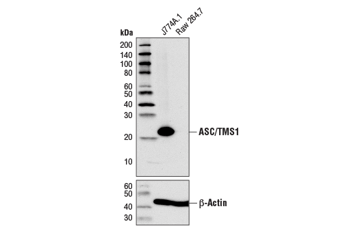
Western blot analysis of extracts from J774A.1 and Raw 264.7 cells using ASC/TMS1 (D2W8U) Rabbit mAb (Mouse Specific) (upper) or β-Actin (D6A8) Rabbit mAb #8457 (lower).
Immunoprecipitation of ASC/TMS1 from J774A.1 cell extracts. Lane 1 is 10% input, lane 2 is Rabbit (DA1E) mAb IgG XP® Isotype Control #3900, and lane 3 is ASC (D2W8U) Rabbit mAb (Mouse Specific). Western blot analysis was performed using ASC/TMS1 (D2W8U) Rabbit mAb (Mouse Specific).
Confocal immunofluorescent analysis of mouse primary bone marrow-derived macrophages (BMDMs) either untreated (upper left) or treated with LPS (50 ng/ml, 4 hr, middle) or LPS followed by ATP (5 mM, 45 min, upper right), and J774A.1 (lower left) or Raw 264.7 (lower right) cells, using ASC/TMS1 (D2W8U) Rabbit mAb (Mouse Specific) (green). Blue pseudocolor = DRAQ5® #4084 (fluorescent DNA dye). Note the translocation of ASC to inflammasomes following stimulation with LPS and ATP (white arrows).
Flow cytometric analysis of Raw264.7 cells (blue) and J774A.1 cells (green) using ASC/TMS1 (D2W8U) Rabbit mAb (Mouse Specific) (solid lines) or a concentration-matched Rabbit (DA1E) mAb IgG XP® Isotype Control #3900 (dashed lines). Anti-rabbit IgG (H+L), F(ab')2 Fragment (Alexa Fluor® 488 Conjugate) #4412 was used as a secondary antibody.

 危险品化学品经营许可证(不带存储) 许可证编号:沪(杨)应急管危经许[2022]202944(QY)
  营业执照(三证合一)