


 全部商品分类
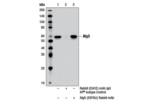


Atg5 (D5F5U) Rabbit mAb


 下载产品说明书 下载COA 下载SDS
 用小程序,查商品更便捷


 收藏
收藏
 对比
对比 咨询
咨询
Monoclonal antibody is produced by immunizing animals with a synthetic peptide corresponding to residues surrounding Leu265 of human Atg5 protein.
Product Usage Information
| Application | Dilution |
|---|---|
| Western Blotting | 1:1000 |
| Simple Western™ | 1:50 - 1:250 |
| Immunoprecipitation | 1:100 |
Specificity/Sensitivity
Species Reactivity:
Human, Mouse, Rat
Supplied in 10 mM sodium HEPES (pH 7.5), 150 mM NaCl, 100 µg/ml BSA, 50% glycerol and less than 0.02% sodium azide. Store at –20°C. Do not aliquot the antibody.
参考图片
Immunoprecipitation of Atg5 from PANC-1 cell extracts using Rabbit (DA1E) mAb IgG XP® Isotype Control #3900 (lane 2) or Atg5 (D5F5U) Rabbit mAb (lane 3). Lane 1 is 10% input. Western blot was performed using Atg5 (D5F5U) Rabbit mAb. Mouse Anti-rabbit IgG (Conformation Specific) (L27A9) mAb #3678 was used as a secondary antibody to avoid cross-reactivity with rabbit IgG.
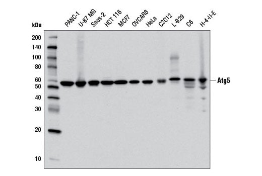
Western blot analysis of extracts from various cell lines using Atg5 (D5F5U) Rabbit mAb.
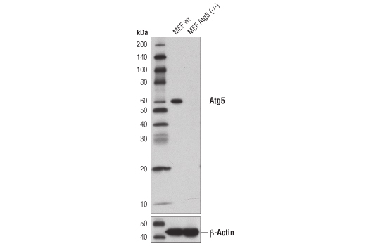
Western blot analysis of extracts from wild-type MEFs (wt) or MEFs from Atg5 knockouts (Atg5-/-) using Atg5 (D5F5U) Rabbit mAb (upper), or β-Actin (D6A8) Rabbit mAb #8457 (lower). Atg5-/- MEFs were kindly provided by Dr. Ramnik Xavier, Massachusetts General Hospital, Harvard Medical School, Boston, MA.
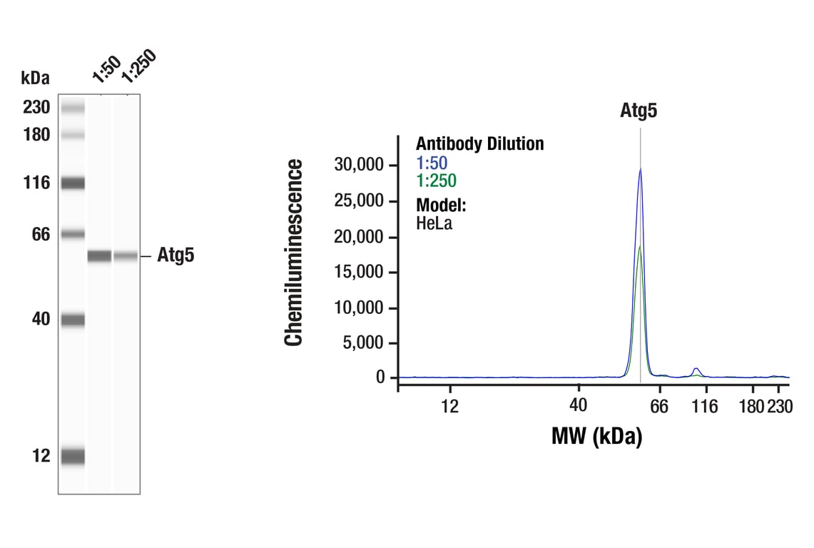
Simple WesternTM analysis of lysates (0.1 mg/mL) from HeLa cells using Atg5 (D5F5U) Rabbit mAb #12994. The virtual lane view (left) shows the target band (as indicated) at 1:50 and 1:250 dilutions of primary antibody. The corresponding electropherogram view (right) plots chemiluminescence by molecular weight along the capillary at 1:50 (blue line) and 1:250 (green line) dilutions of primary antibody. This experiment was performed under reducing conditions on the JessTM Simple Western instrument from ProteinSimple, a BioTechne brand, using the 12-230 kDa separation module.