

 全部商品分类
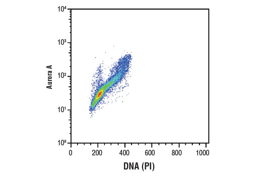


 用小程序,查商品更便捷


Monoclonal antibody is produced by immunizing animals with a synthetic peptide corresponding to residues surrounding Pro70 of human Aurora A protein.
Product Usage Information
| Application | Dilution |
|---|---|
| Western Blotting | 1:1000 |
| Immunoprecipitation | 1:50 |
| Immunofluorescence (Immunocytochemistry) | 1:100 - 1:200 |
| Flow Cytometry (Fixed/Permeabilized) | 1:50 - 1:200 |
Specificity/Sensitivity
Species Reactivity:
Human
Supplied in 10 mM sodium HEPES (pH 7.5), 150 mM NaCl, 100 µg/ml BSA, 50% glycerol and less than 0.02% sodium azide. Store at –20°C. Do not aliquot the antibody.
For a carrier free (BSA and azide free) version of this product see product #79037.
参考图片
Flow cytometric analysis of Jurkat cells using Aurora A (D3E4Q) Rabbit mAb and Propidium Iodide (PI)/RNase Staining Solution #4087 to measure DNA content. Anti-rabbit IgG (H+L), F(ab')2 Fragment (Alexa Fluor® 488 Conjugate) #4412 was used as a secondary antibody.
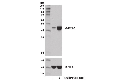
Western blot analysis of extracts from HT-29 cells, untreated (-) or synchronized in mitosis by treatment with thymidine (2 mM, 17 hr; +) and Nocodazole #2190 (100 ng/ml, 24 hr; +), using Aurora A (D3E4Q) Rabbit mAb (upper) and β-Actin (D6A8) Rabbit mAb #8457 (lower).
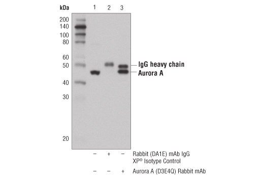
Immunoprecipitation of Aurora A from HeLa cell extracts using Rabbit (Da1E) mAb IgG XP® Isotype Control #3900 (lane 2) or Aurora A (D3E4Q) Rabbit mAb (lane 3). Lane 1 is 10% input. Western blot analysis was performed using Aurora A (D3E4Q) Rabbit mAb.
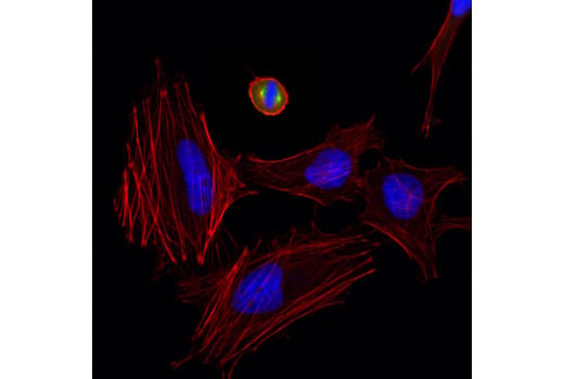
Confocal immunofluorescent analysis of HeLa cells using Aurora A (D3E4Q) Rabbit mAb (green). Actin filaments were labeled with DyLight™ 554 Phalloidin #13054 (red). Blue pseudocolor = DRAQ5® #4084 (fluorescent DNA dye).