


 全部商品分类
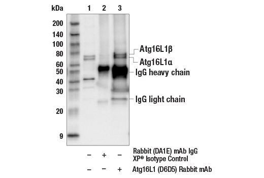


Autophagy Antibody Sampler Kit

 下载产品说明书 下载SDS
 用小程序,查商品更便捷


 收藏
收藏
 对比
对比 咨询
咨询
The Autophagy Antibody Sampler Kit provides an economical means to investigate the molecular machinery of autophagy within the cell. The kit contains enough primary and secondary antibodies to perform two Western blot experiments.
参考图片
Immunoprecipitation of Atg16L1 from Jurkat cell extracts. Lane 1 is 10% input, lane 2 is precipitated with Rabbit (DA1E) mAb IgG XP® Isotype Control #3900, and lane 3 is Atg16L1 (D6D5) Rabbit mAb, #8089. Western blot was performed using Atg16L1 (D6D5) Rabbit mAb.
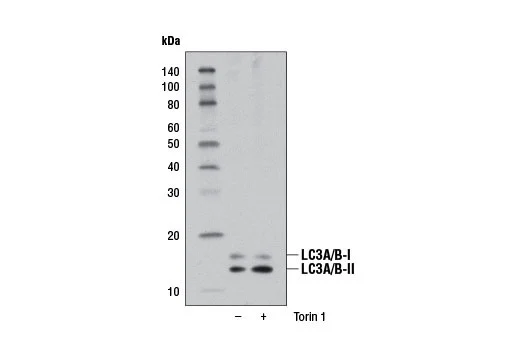
Western blot analysis of extracts from RD cells, untreated (-) or Torin 1-treated (250 nM, 4 hr; +), using LC3A/B (D3U4C) XP® Rabbit mAb.
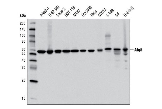
Western blot analysis of extracts from various cell lines using Atg5 (D5F5U) Rabbit mAb.
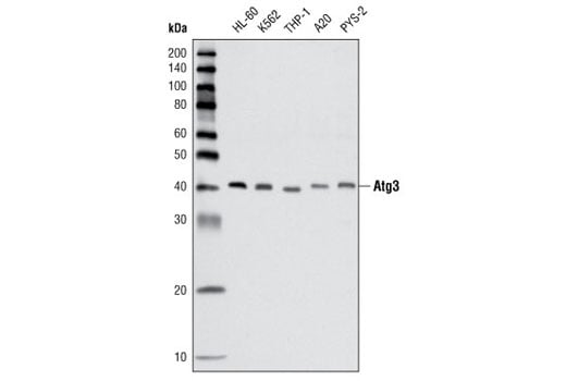
Western blot analysis of extracts from various cell lines using Atg3 Antibody.
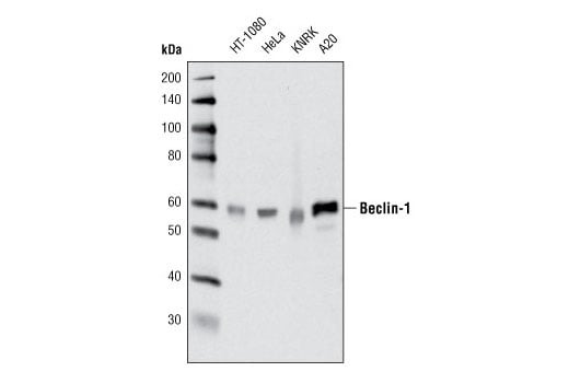
Western blot analysis of extracts from various cell lines using Beclin-1 (D40C5) Rabbit mAb.
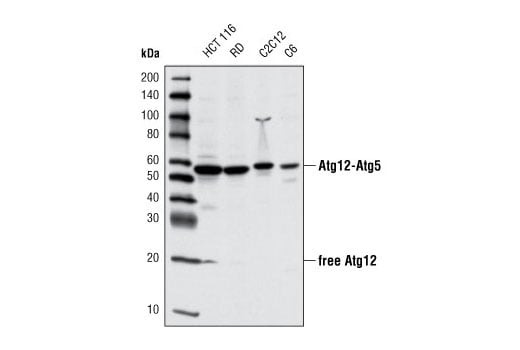
Western blot analysis of extracts from various cell lines using Atg12 (D88H11) Rabbit mAb.
After the primary antibody is bound to the target protein, a complex with HRP-linked secondary antibody is formed. The LumiGLO® is added and emits light during enzyme catalyzed decomposition.
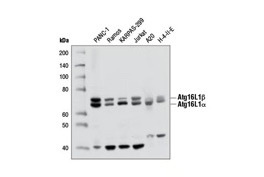
Western blot analysis of extracts from various cell lines using Atg16L1 (D6D5) Rabbit mAb.
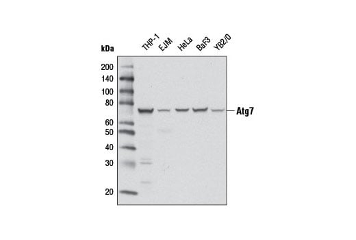
Western blot analysis of extracts from various cell lines using Atg7 (D12B11) Rabbit mAb.
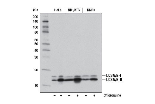
Western blot analysis of extracts from HeLa, NIH/3T3, and KNRK cells, untreated (-) or chloroquine-treated (50 μM, overnight; +), using LC3A/B (D3U4C) XP® Rabbit mAb.
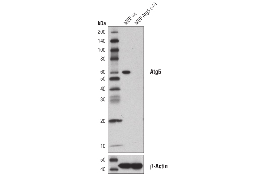
Western blot analysis of extracts from wild-type MEFs (wt) or MEFs from Atg5 knockouts (Atg5-/-) using Atg5 (D5F5U) Rabbit mAb (upper), or β-Actin (D6A8) Rabbit mAb #8457 (lower). Atg5-/- MEFs were kindly provided by Dr. Ramnik Xavier, Massachusetts General Hospital, Harvard Medical School, Boston, MA.
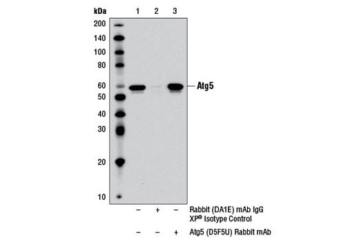
Immunoprecipitation of Atg5 from PANC-1 cell extracts using Rabbit (DA1E) mAb IgG XP® Isotype Control #3900 (lane 2) or Atg5 (D5F5U) Rabbit mAb (lane 3). Lane 1 is 10% input. Western blot was performed using Atg5 (D5F5U) Rabbit mAb. Mouse Anti-rabbit IgG (Conformation Specific) (L27A9) mAb #3678 was used as a secondary antibody to avoid cross-reactivity with rabbit IgG.
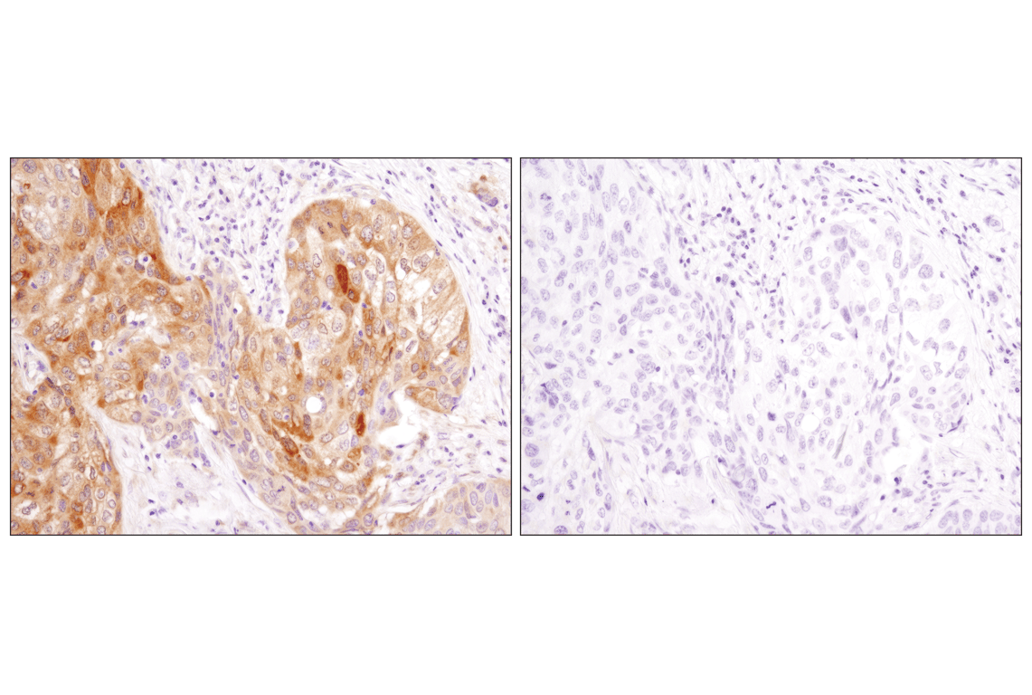
Immunohistochemical analysis of paraffin-embedded human squamous cell lung carcinoma using LC3A/B (D3U4C) XP® Rabbit mAb in the presence of control peptide (left) or antigen-specific peptide (right).
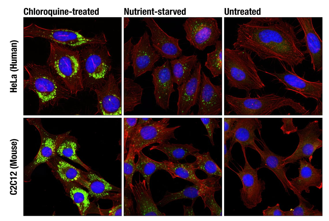
Confocal immunofluorescent analysis of HeLa (upper) and C2C12 (lower) cells, chloroquine-treated (50 μM, overnight; left), nutrient-starved with EBSS (3 hr, middle) or untreated (right) using LC3A/B (D3U4C) XP® Rabbit mAb (green) and β-Actin (13E5) Rabbit mAb (Alexa Fluor® 555 Conjugate) #8046 (red). Blue pseudocolor= DRAQ5® #4084 (fluorescent DNA dye).
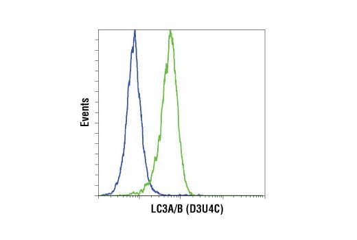
Flow cytometric analysis of HeLa cells, untreated (blue) or treated with chloroquine (50 µM, 16 hr; green) using LC3A/B (D3U4C) Rabbit mAb. Anti-rabbit IgG (H+L), F(ab')2 Fragment (Alexa Fluor® 647 Conjugate) #4414 was used as a secondary antibody.
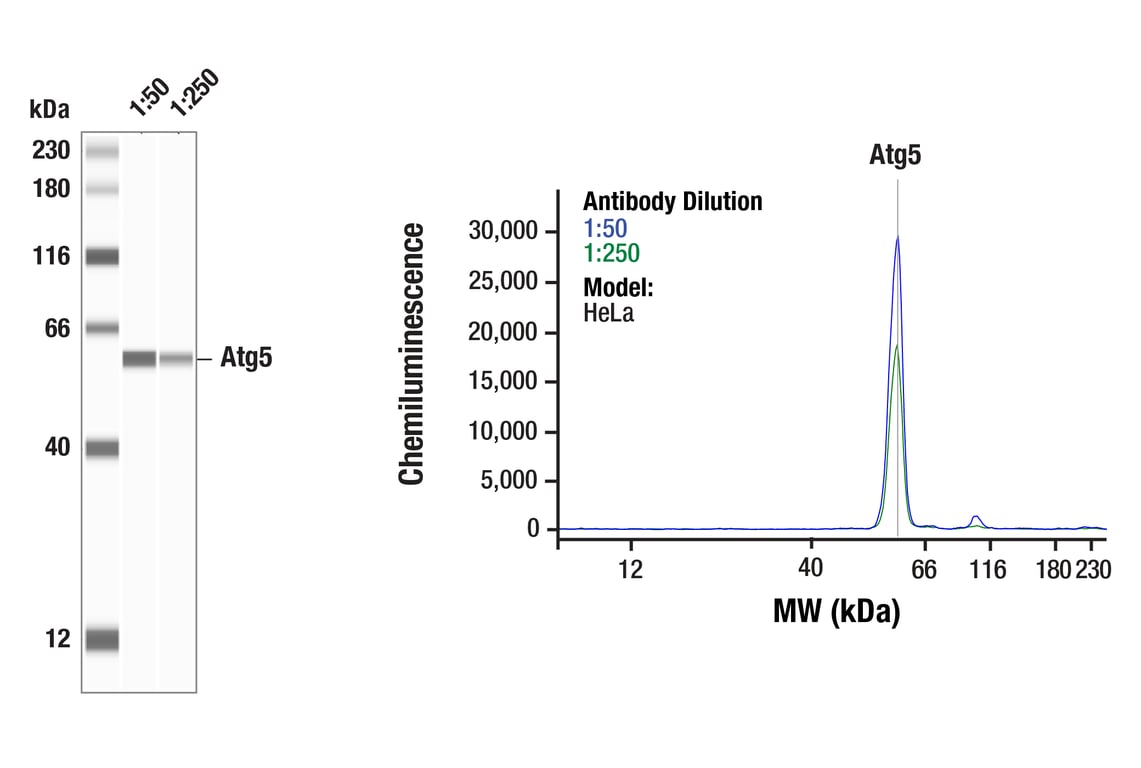
Simple WesternTM analysis of lysates (0.1 mg/mL) from HeLa cells using Atg5 (D5F5U) Rabbit mAb #12994. The virtual lane view (left) shows the target band (as indicated) at 1:50 and 1:250 dilutions of primary antibody. The corresponding electropherogram view (right) plots chemiluminescence by molecular weight along the capillary at 1:50 (blue line) and 1:250 (green line) dilutions of primary antibody. This experiment was performed under reducing conditions on the JessTM Simple Western instrument from ProteinSimple, a BioTechne brand, using the 12-230 kDa separation module.
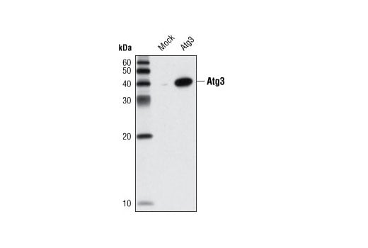
Western blot analysis of extracts from HeLa cells, mock transfected or transfected with mouse Atg3, using Atg3 Antibody.
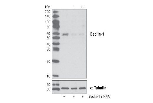
Western blot analysis of extracts from HeLa cells, transfected with 100 nM SignalSilence® Control siRNA (Unconjugated) #6568 (-), SignalSilence® Beclin-1 siRNA I #6222 (+) or SignalSilence® Beclin-1 siRNA II (+), using Beclin-1 (D40C5) XP® Rabbit mAb #3495 (upper) or α-Tubulin (11H10) Rabbit mAb #2125 (lower). The Beclin-1 (D40C5) XP® Rabbit mAb confirms silencing of Beclin-1 expression, while the α-Tubulin (11H10) Rabbit mAb is used to control for loading and specificity of Beclin-1 siRNA.
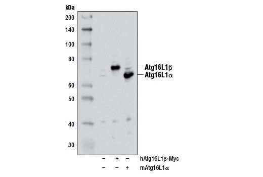
Western blot analysis of extracts from 293T cells, mock transfected (-) or transfected with either a myc-tagged human Atg16L1β construct (hAtg16L1β-Myc; +) or a mouse Atg16L1α construct (mAtg16L1α; +), using Atg16L1 (D6D5) Rabbit mAb. The myc-tagged human Atg16L1β construct was kindly provided by Dr. Qing Zhong, University of California Berkeley.
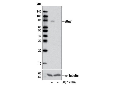
Western blot analysis of extracts from HeLa cells, transfected with 100 nM SignalSilence® Control siRNA (Unconjugated) #6568 (-) or SignalSilence® Atg7 siRNA I #6604 (+), using Atg7 (D12B11) Rabbit mAb (upper) or α-Tubulin (11Η10) Rabbit mAb #2125 (lower). The Atg7 (D12B11) Rabbit mAb confirms silencing of Atg7 expression, while the α-Tubulin (11H10) Rabbit mAb is used as a loading control.
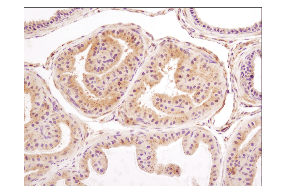
Immunohistochemical analysis of paraffin-embedded mouse prostate using LC3A/B (D3U4C) XP® Rabbit mAb.
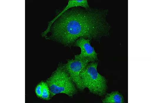
Confocal immunofluorescent analysis of EBSS-starved PANC-1 cells using Atg16L1 (D6D5) Rabbit mAb (green). Blue pseudocolor = DRAQ5® #4084 (fluorescent DNA dye).
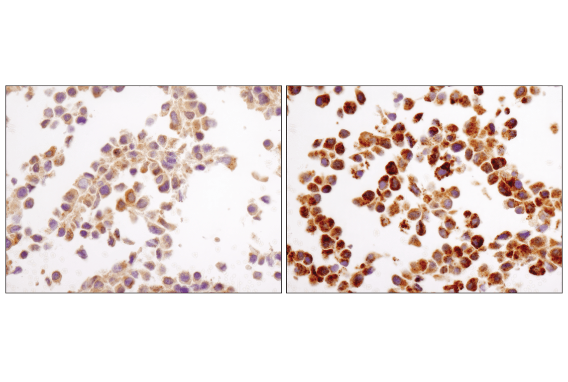
Immunohistochemical analysis of paraffin-embedded NIH/3T3 cell pellets, control (left) or chloroquine-treated (right), using LC3A/B (D3U4C) XP® Rabbit mAb.