


 全部商品分类


 用小程序,查商品更便捷


Monoclonal antibody is produced by immunizing animals with a synthetic peptide corresponding to residues surrounding Gly47 of human Bcl-2 protein.
Product Usage Information
| Application | Dilution |
|---|---|
| Western Blotting | 1:1000 |
| Simple Western™ | 1:10 - 1:50 |
| Immunoprecipitation | 1:50 |
| IHC Leica Bond | 1:800 - 1:3200 |
| Immunohistochemistry (Paraffin) | 1:400 - 1:1600 |
| Flow Cytometry (Fixed/Permeabilized) | 1:200 - 1:800 |
Specificity/Sensitivity
Species Reactivity:
Human
Supplied in 10 mM sodium HEPES (pH 7.5), 150 mM NaCl, 100 µg/ml BSA, 50% glycerol and less than 0.02% sodium azide. Store at –20°C. Do not aliquot the antibody.
For a carrier-free (BSA and azide free) version of this product see product #17447
参考图片
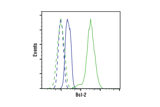
Flow cytometric analysis of HT-29 cells (blue) and RL cells (green) using Bcl-2 (124) Mouse mAb (solid lines) or a concentration-matched Mouse (G3A1) mAb IgG1 Isotype Control #5415 (dashed lines). Anti-mouse IgG (H+L), F(ab')2 Fragment (Alexa Fluor® 488 Conjugate) #4408 was used as a secondary antibody.
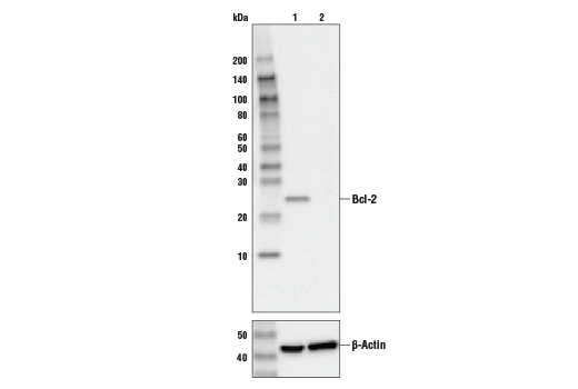
Western blot analysis of extracts from control HeLa cells (lane 1) or Bcl-2 knockout HeLa cells (lane 2) using Bcl-2 (124) Mouse mAb #15071 (upper), or β-actin (13E5) Rabbit mAb #4970 (lower). The absence of signal in the Bcl-2-knockout HeLa cells confirms specificity of the antibody for Bcl-2.
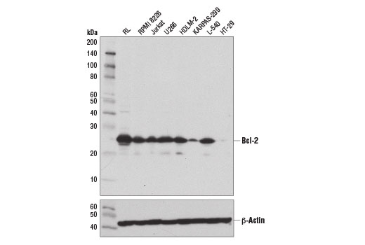
Western blot analysis of extracts from various cell lines using Bcl-2 (124) Mouse mAb (upper) and β-Actin (D6A8) Rabbit mAb #8457 (lower). KARPAS cell Line source: Dr Abraham Karpas at the University of Cambridge.
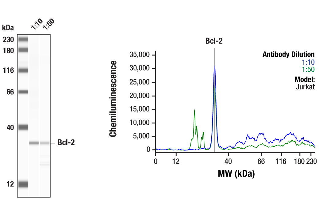
Simple Western™ analysis of lysates (1 mg/mL) from Jurkat cells using Bcl-2 (124) Mouse mAb #15071. The virtual lane view (left) shows the target band (as indicated) at 1:10 and 1:50 dilutions of primary antibody. The corresponding electropherogram view (right) plots chemiluminescence by molecular weight along the capillary at 1:10 (blue line) and 1:50 (green line) dilutions of primary antibody. This experiment was performed under reducing conditions on the Jess™ Simple Western instrument from ProteinSimple, a BioTechne brand, using the 12-230 kDa separation module.
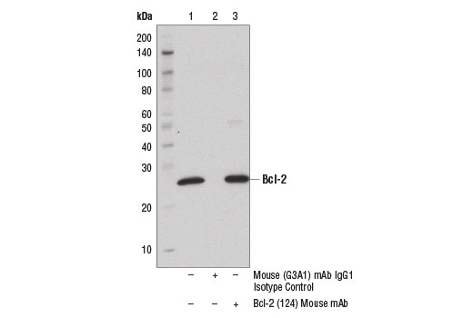
Immunoprecipitation of Bcl-2 from RL cell extracts using Mouse (G3A1) mAb IgG1 Isotype Control #5415 (lane 2) or Bcl-2 (124) Mouse mAb (lane 3). Lane 1 is 10% input. Western blot was performed using Bcl-2 (D55G8) Rabbit mAb (Human Specific) #4223.
Immunohistochemical analysis of paraffin-embedded human ductal breast carcinoma using Bcl-2 (124) mouse mAb performed on the Leica® BOND™ Rx.
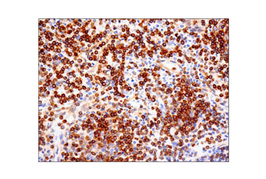
Immunohistochemical analysis of paraffin-embedded human non-Hodgkin’s lymphoma using Bcl-2 (124) mouse mAb performed on the Leica® BOND™ Rx.
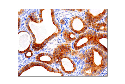
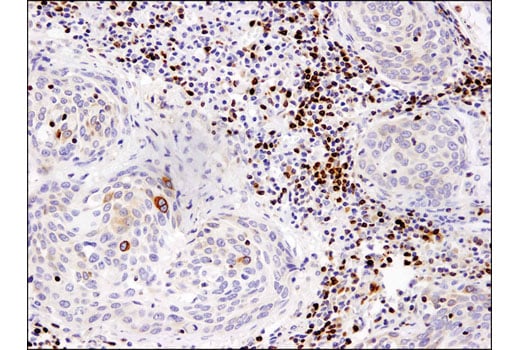
Immunohistochemical analysis of paraffin-embedded human ovarian clear cell carcinoma using Bcl-2 (124) mouse mAb performed on the Leica® BOND™ Rx.
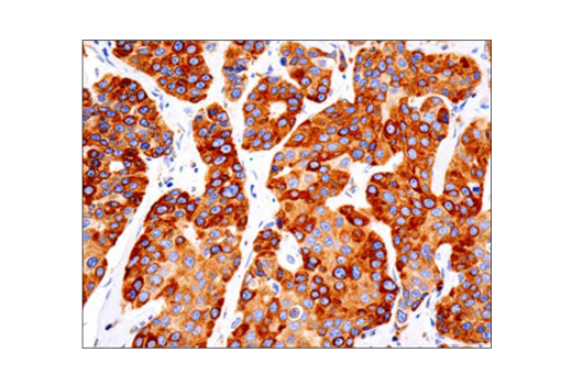
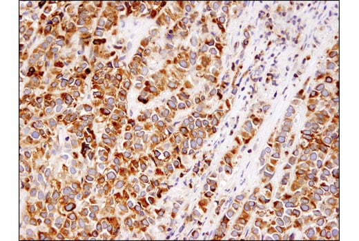
Immunohistochemical analysis of paraffin-embedded human breast carcinoma using Bcl-2 (124) Mouse mAb.
Immunohistochemical analysis of paraffin-embedded human lung carcinoma using Bcl-2 (124) Mouse mAb.
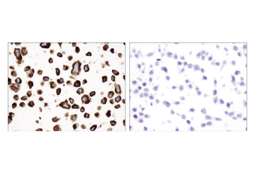
Immunohistochemical analysis of paraffin-embedded RL (positive, left) and HT-29 (negative, right) cell pellets using Bcl-2 (124) Mouse mAb.
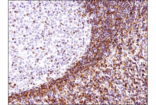
Immunohistochemical analysis of paraffin-embedded human tonsil using Bcl-2 (124) Mouse mAb.