


 全部商品分类
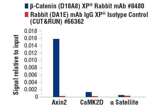


 用小程序,查商品更便捷


Monoclonal antibody is produced by immunizing animals with a synthetic peptide corresponding to residues surrounding Pro714 of human β-catenin protein.
Product Usage Information
For optimal ChIP and ChIP-seq results, use 20 μl of antibody and 10 μg of chromatin (approximately 4 x 10⁶ cells) per IP. This antibody has been validated using SimpleChIP® Enzymatic Chromatin IP Kits.
The CUT&RUN dilution was determined using CUT&RUN Assay Kit #86652.
| Application | Dilution |
|---|---|
| Western Blotting | 1:1000 |
| Simple Western™ | 1:10 - 1:50 |
| Immunoprecipitation | 1:50 |
| IHC Leica Bond | 1:50 - 1:200 |
| Immunohistochemistry (Paraffin) | 1:50 - 1:200 |
| Immunofluorescence (Frozen) | 1:50 - 1:100 |
| Immunofluorescence (Immunocytochemistry) | 1:100 - 1:400 |
| Flow Cytometry (Fixed/Permeabilized) | 1:50 - 1:100 |
| Chromatin IP | 1:25 |
| Chromatin IP-seq | 1:25 |
| CUT&RUN | 1:25 |
Specificity/Sensitivity
Species Reactivity:
Human, Mouse, Rat, Monkey
Supplied in 10 mM sodium HEPES (pH 7.5), 150 mM NaCl, 100 µg/ml BSA, 50% glycerol and less than 0.02% sodium azide. Store at –20°C. Do not aliquot the antibody.
For a carrier free (BSA and azide free) version of this product see product #84441.
参考图片
CUT&RUN was performed with HCT 116 cells and either β-Catenin (D10A8) XP® Rabbit mAb or Rabbit (DA1E) mAb IgG XP® Isotype Control (CUT&RUN) #66362, using CUT&RUN Assay Kit #86652. The enriched DNA was quantified by real-time PCR using SimpleChIP® Human Axin2 Intron 1 Primers #8973, SimpleChIP® Human CaMK2D Intron 3 Primers #5111 and SimpleChIP® Human α Satellite Repeat Primers #4486. The amount of immunoprecipitated DNA in each sample is represented as signal relative to the total amount of input chromatin, which is equivalent to one.
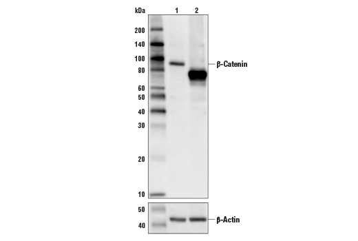
Western blot analysis of extracts from control HeLa cells (lane 1) or HeLa cells with an apparent in-frame truncation mutation in the gene encoding β-Catenin (lane 2) using β-Catenin (D10A8) XP® Rabbit mAb, #8480 (upper) or β-actin (D6A8) Rabbit mAb #8457 (lower). The change in β-Catenin molecular weight in the mutated HeLa cells is consistent with an in-frame deletion.
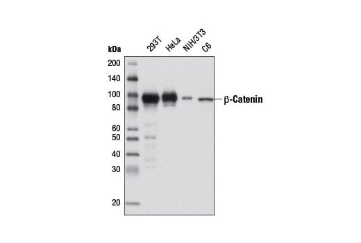
Western blot analysis of extracts from various cell lines using β-Catenin (D10A8) XP® Rabbit mAb.
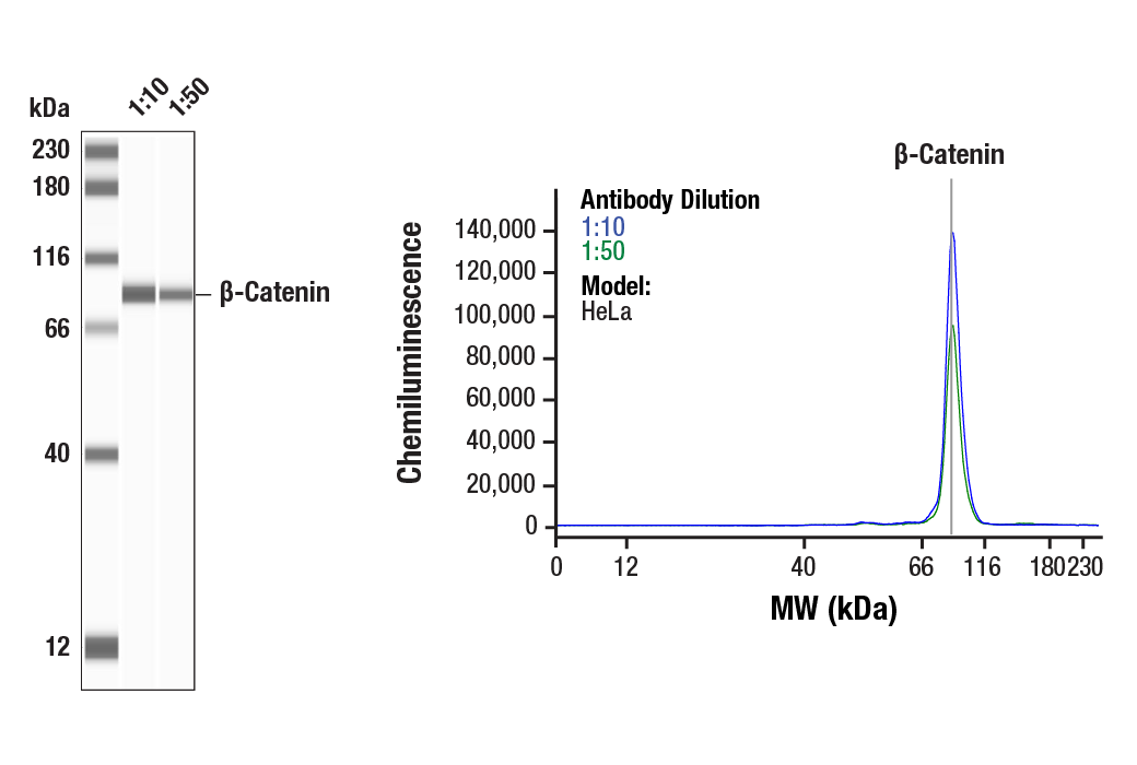
Simple Western™ analysis of lysates (0.1 mg/mL) from HeLa cells using β-Catenin (D10A8) XP® Rabbit mAb #8480. The virtual lane view (left) shows the target band (as indicated) at 1:10 and 1:50 dilutions of primary antibody. The corresponding electropherogram view (right) plots chemiluminescence by molecular weight along the capillary at 1:10 (blue line) and 1:50 (green line) dilutions of primary antibody. This experiment was performed under reducing conditions on the Jess™ Simple Western instrument from ProteinSimple, a BioTechne brand, using the 12-230 kDa separation module.
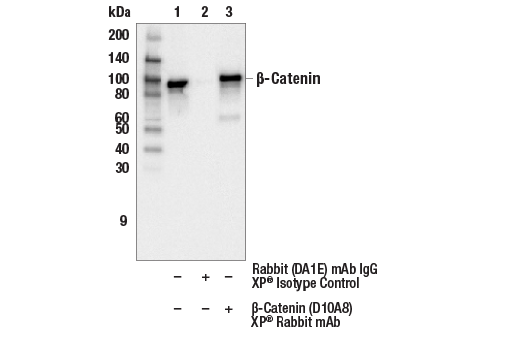
Immunoprecipitation of β-Catenin from HeLa cell extracts. Lane 1 is 10% input, lane 2 is precipitated with Rabbit (DA1E) mAb IgG XP® Isotype Control #3900, and lane 3 is β-Catenin (D10A8) XP® Rabbit mAb, #8480. Western blot was performed using β-Catenin (15B8) Mouse mAb, #37447.
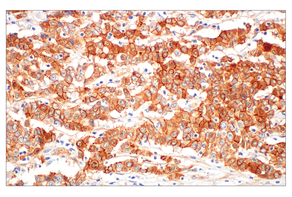
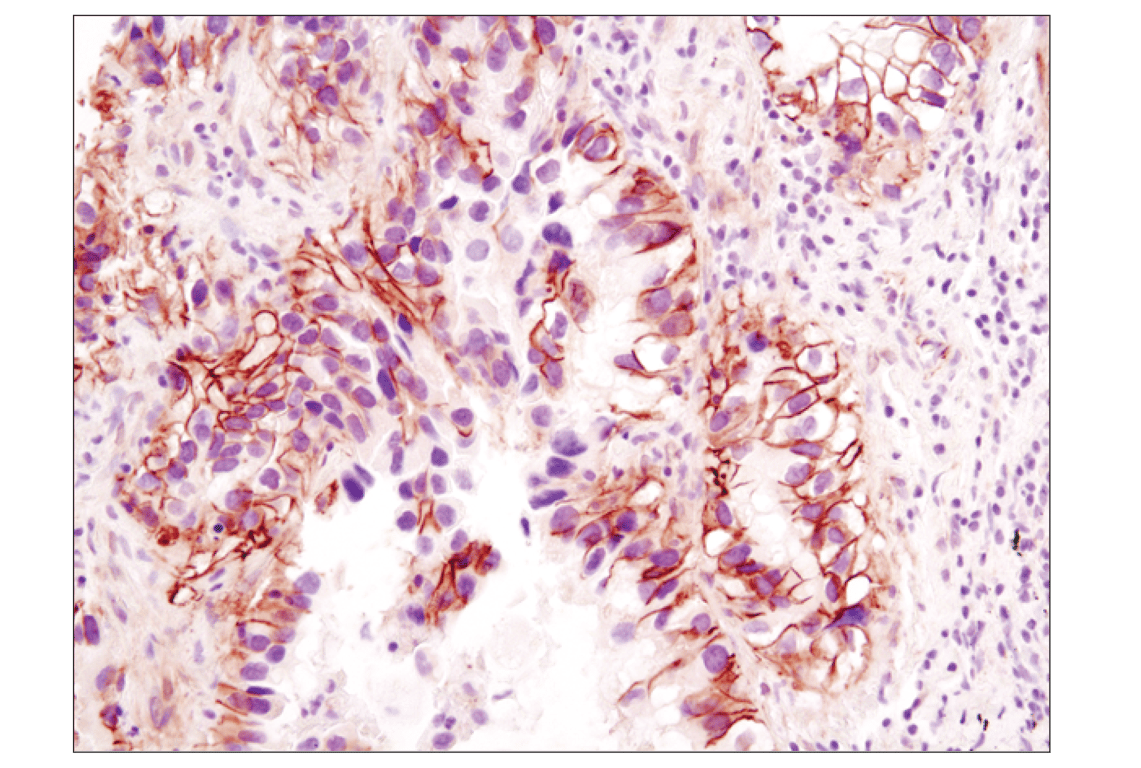
Immunohistochemical analysis of paraffin-embedded human prostate adenocarcinoma using ß-Catenin (D10A8) XP® Rabbit mAb performed on the Leica® BOND™ Rx.
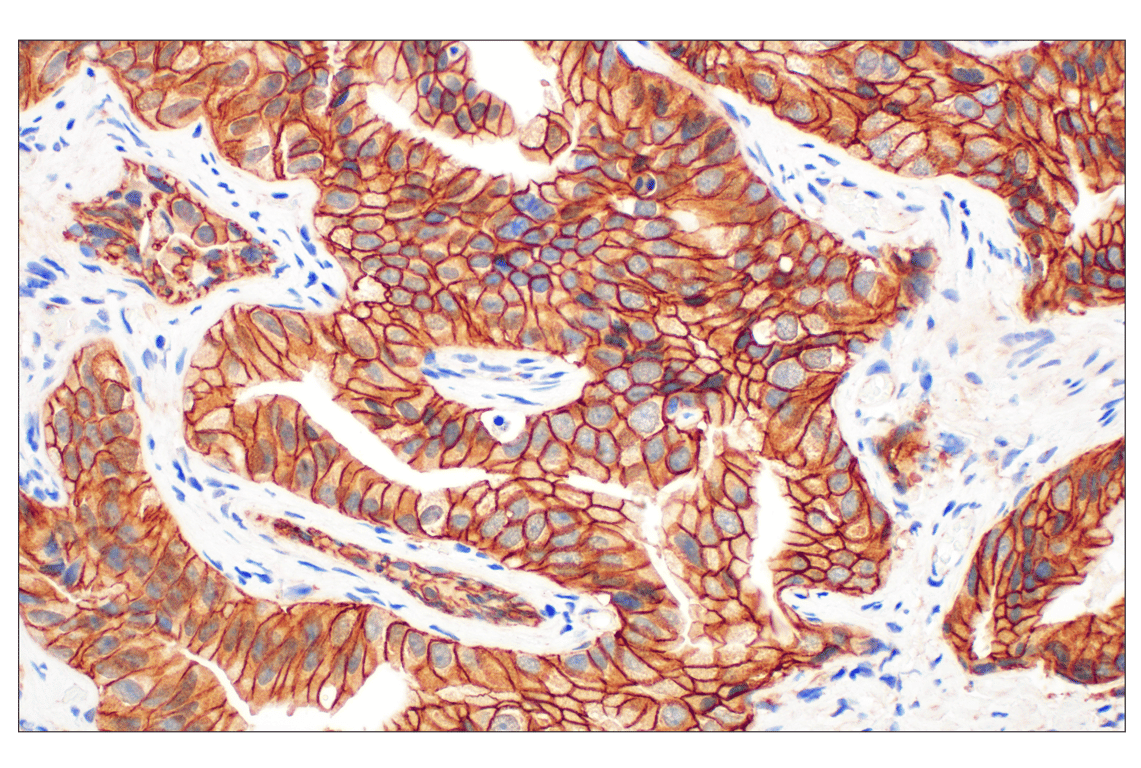
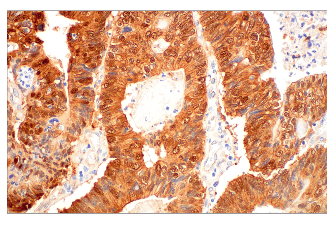
Immunohistochemical analysis of paraffin-embedded human colon adenocarcinoma using ß-Catenin (D10A8) XP® Rabbit mAb performed on the Leica® BOND™ Rx.
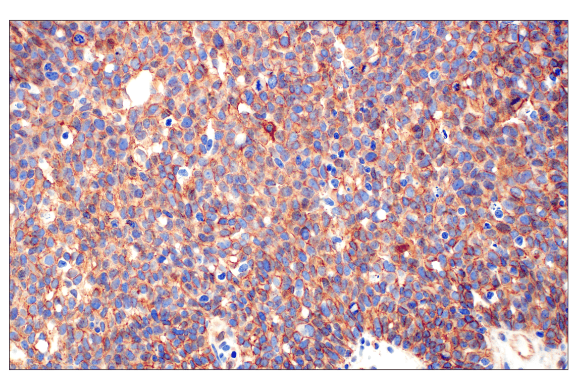
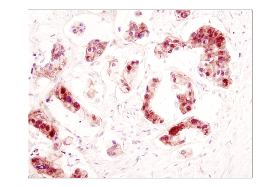
Immunohistochemical analysis of paraffin-embedded human serous adenocarcinoma of the ovary using ß-Catenin (D10A8) XP® Rabbit mAb performed on the Leica® BOND™ Rx.
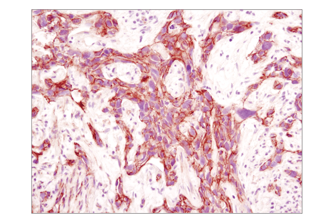
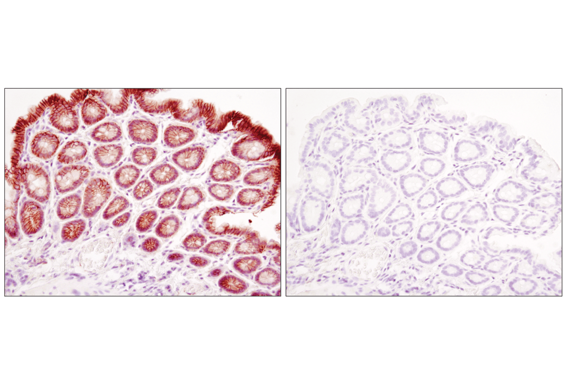
Immunohistochemical analysis of paraffin-embedded human colon adenocarcinoma using ß-Catenin (D10A8) XP® Rabbit mAb.
Immunohistochemical analysis of paraffin-embedded human lung carcinoma using β-Catenin (D10A8) XP® Rabbit mAb.
Immunohistochemical analysis of paraffin-embedded human colon adenocarcinoma using ß-Catenin (D10A8) XP® Rabbit mAb.
Immunohistochemical analysis of paraffin-embedded human colon carcinoma using β-Catenin (D10A8) XP® Rabbit mAb.
Immunohistochemical analysis of paraffin-embedded human breast carcinoma using β-Catenin (D10A8) XP® Rabbit mAb.
Immunohistochemical analysis of paraffin-embedded mouse colon using β-Catenin (D10A8) XP® Rabbit mAb in the presence of control peptide (left) or antigen-specific peptide (right).
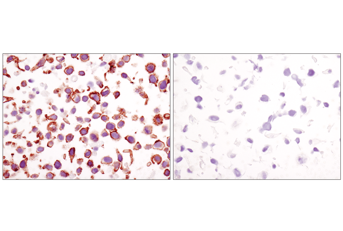
Immunohistochemical analysis of paraffin-embedded cell pellets, HeLa (left) or NCI-H28 (right), using β-Catenin (D10A8) XP® Rabbit mAb.
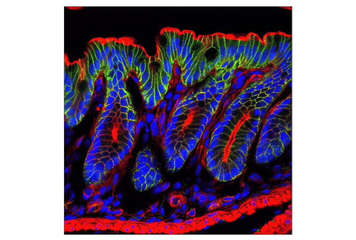
Confocal immunofluorescent analysis of mouse colon using β-Catenin (D10A8) XP® Rabbit mAb (green). Actin filaments were labeled with DY-554 phalloidin (red). Blue pseudocolor = DRAQ5® #4084 (fluorescent DNA dye).
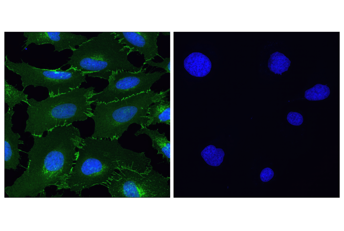
Confocal immunofluorescent analysis of HeLa (left) and NCI-H28 (right) cells using β-Catenin (D10A8) XP® Rabbit mAb (green). Blue pseudocolor = DRAQ5® #4084 (fluorescent DNA dye).
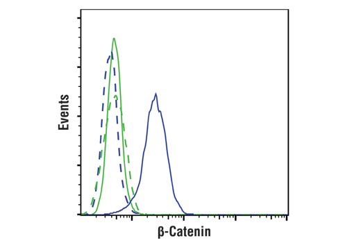
Flow cytometric analysis of NCI-H28 cells (green) and HeLa cells (blue) using β-Catenin (D10A8) XP® Rabbit mAb (solid lines) or concentration-matched Rabbit Isotype Control #3900 (dashed lines). Anti-rabbit IgG (H+L), F(ab')2 Fragment (Alexa Fluor® 488 Conjugate) #4412 was used as secondary antibody.
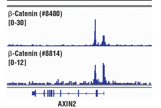
Chromatin immunoprecipitations were performed with cross-linked chromatin from HCT116 cells and either β-Catenin (D10A8) XP® Rabbit mAb or Non-phospho (Active) β-Catenin (Ser33/37/Thr41) (D13A1) Rabbit mAb #8814, using SimpleChIP® Enzymatic Chromatin IP Kit (Magnetic Beads) #9005. DNA Libraries were prepared using DNA Library Prep Kit for Illumina® (ChIP-seq, CUT&RUN) #56795. The figure shows binding across AXIN2, a known target gene of β-Catenin (see additional figure containing ChIP-qPCR data).
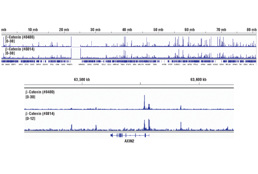
Chromatin immunoprecipitations were performed with cross-linked chromatin from HCT116 cells and either β-Catenin (D10A8) XP® Rabbit mAb or Non-phospho (Active) β-Catenin (Ser33/37/Thr41) (D13A1) Rabbit mAb #8814, using SimpleChIP® Enzymatic Chromatin IP Kit (Magnetic Beads) #9005. DNA Libraries were prepared using DNA Library Prep Kit for Illumina® (ChIP-seq, CUT&RUN) #56795. The figure shows binding across chromosome 17 (upper), including AXIN2 (lower), a known target gene of β-Catenin (see additional figure containing ChIP-qPCR data).
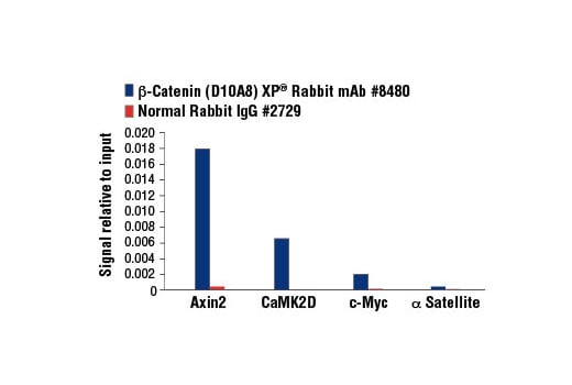
Chromatin immunoprecipitations were performed with cross-linked chromatin from HCT 116 cells and either β-Catenin (D10A8) XP® Rabbit mAb or Normal Rabbit IgG #2729 using SimpleChIP® Enzymatic Chromatin IP Kit (Magnetic Beads) #9003. The enriched DNA was quantified by real-time PCR using SimpleChIP® Human Axin2 Intron 1 Primers #8973, SimpleChIP® Human CaMK2D Intron 3 Primers #5111, human c-Myc promoter primers, and SimpleChIP® Human α Satellite Repeat Primers #4486. The amount of immunoprecipitated DNA in each sample is represented as signal relative to the total amount of input chromatin, which is equivalent to one.
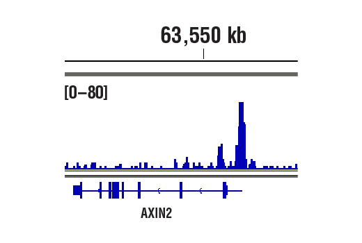
CUT&RUN was performed with HCT 116 cells and β-Catenin (D10A8) XP® Rabbit mAb, using CUT&RUN Assay Kit #86652. DNA library was prepared using DNA Library Prep Kit for Illumina® (ChIP-seq, CUT&RUN) #56795. The figure shows binding across Axin2, a known target gene of β-Catenin (see additional figure containing CUT&RUN-qPCR data).
CUT&RUN was performed with HCT 116 cells and β-Catenin (D10A8) XP® Rabbit mAb, using CUT&RUN Assay Kit #86652. DNA Libraries were prepared using DNA Library Prep Kit for Illumina® (ChIP-seq, CUT&RUN) #56795. The figures show binding across chromosome 17 (upper), including Axin2 (lower), a known target gene of β-Catenin (see additional figure containing CUT&RUN-qPCR data).