


 全部商品分类
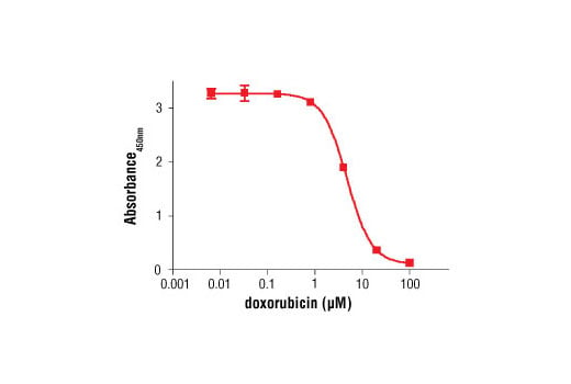


 用小程序,查商品更便捷


The BrdU Cell Proliferation Assay Kit detects 5-bromo-2’-deoxyuridine (BrdU) incorporated into cellular DNA during cell proliferation using an anti-BrdU antibody. When cells are cultured with labeling medium that contains BrdU, this pyrimidine analog is incorporated in place of thymidine into the newly synthesized DNA of proliferating cells. After removing labeling medium, cells are fixed and the DNA is denatured with our fixing/denaturing solution. Then a BrdU mouse mAb is added to detect the incorporated BrdU (The denaturing of DNA is necessary to improve the accessibility of the incorporated BrdU to the detection antibody). Anti-mouse IgG, HRP-linked antibody is then used to recognize the bound detection antibody. HRP substrate TMB is added to develop color. The magnitude of the absorbance for the developed color is proportional to the quantity of BrdU incorporated into cells, which is a direct indication of cell proliferation.

Specificity/Sensitivity
参考图片
Figure 3. Jurkat cells were seeded at 4x10⁴ cells/well in a 96-well plate and incubated overnight. Cells were then treated with various concentrations of doxorubicin for 2 hr. Finally, 10 μM BrdU was added to the plate and cells were incubated for 4 hr.
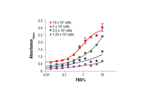
Figure 1. C2C12 cells were seeded at varying density in serum free medium in a 96-well plate and incubated overnight. Serum was added to the plate at various concentrations and cells were incubated for 24 hr. Finally, 10 μM BrdU was added to the plate and cells were incubated for 4 hr.
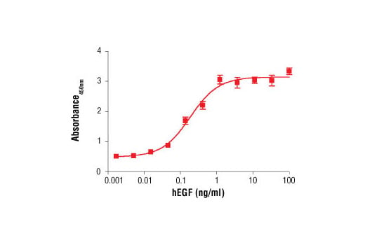
Figure 2. Treatment of MCF 10A cells with Human Epidermal Growth Factor (hEGF) #8916 increases cell proliferation as detected by BrdU Cell Proliferation Assay Kit #6813. MCF 10A cells were seeded at 1x10⁴ cells/well in a 96-well plate and incubated overnight. Cells were then starved in serum free medium overnight. hEGF was added to the plate and cells were incubated for 24 hr. Finally, 10 μM BrdU was added to the plate and cells were incubated for 4 hr.