


 全部商品分类
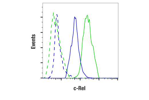


 用小程序,查商品更便捷


Monoclonal antibody is produced by immunizing animals with a synthetic peptide corresponding to residues near the carboxy terminus of human c-Rel protein.

Product Usage Information
| Application | Dilution |
|---|---|
| Western Blotting | 1:1000 |
| Immunoprecipitation | 1:200 |
| Immunohistochemistry (Paraffin) | 1:50 - 1:200 |
| Immunofluorescence (Immunocytochemistry) | 1:50 - 1:200 |
| Flow Cytometry (Fixed/Permeabilized) | 1:200 - 1:800 |
Specificity/Sensitivity
Species Reactivity:
Human
Supplied in 10 mM sodium HEPES (pH 7.5), 150 mM NaCl, 100 µg/mL BSA, 50% glycerol, and less than 0.02% sodium azide. Store at –20°C. Do not aliquot the antibody.
For a carrier free (BSA and azide free) version of this product see product #57603.
参考图片
Flow cytometric analysis of HL-60 cells (blue, low-expressing) and RT-4 cells (green, high-expressing) using c-Rel (E8Z5Y) XP® Rabbit mAb (solid lines) or concentration-matched Rabbit (DA1E) mAb IgG XP® Isotype Control #3900 (dashed lines). Anti-rabbit IgG (H+L), F(ab')2 Fragment (Alexa Fluor® 488 Conjugate) #4412 was used as a secondary antibody.
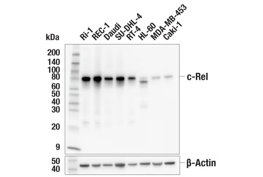
Western blot analysis of extracts from various cell lines using c-Rel (E8Z5Y) XP® Rabbit mAb (upper) or β-Actin (D6A8) Rabbit mAb #8457 (lower). Lower expression of c-Rel protein in HL-60, MDA-MB-453, and Caki-1 cells is consistent with the predicted expression pattern.
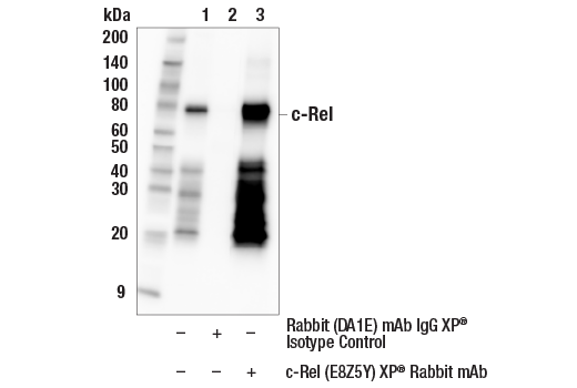
Immunoprecipitation of c-Rel protein from RT-4 cell extracts. Lane 1 is 10% input, lane 2 is Rabbit (DA1E) mAb IgG XP® Isotype Control #3900, and lane 3 is c-Rel (E8Z5Y) XP® Rabbit mAb. Western blot analysis was performed using c-Rel (E8Z5Y) XP® Rabbit mAb. Mouse Anti-rabbit IgG (Conformation Specific) (L27A9) mAb (HRP Conjugate) #5127 was used as a secondary antibody.
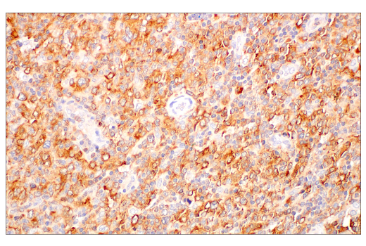
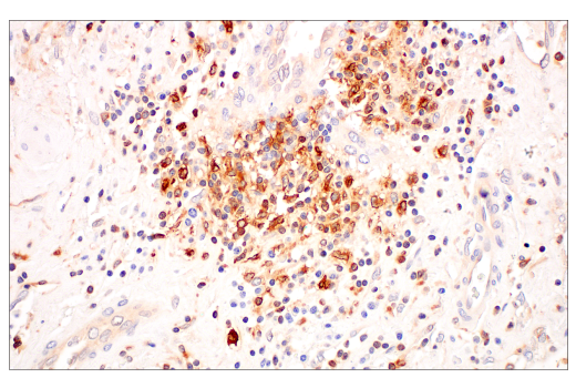
Immunohistochemical analysis of paraffin-embedded human T-cell lymphoma using c-Rel (E8Z5Y) XP® Rabbit mAb.
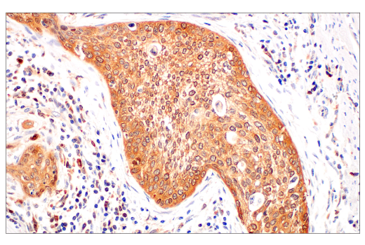
Immunohistochemical analysis of paraffin-embedded human squamous cell carcinoma of the cervix using c-Rel (E8Z5Y) XP® Rabbit mAb.
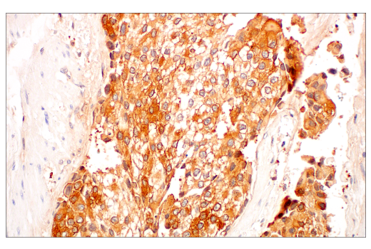
Immunohistochemical analysis of paraffin-embedded human urothelial carcinoma using c-Rel (E8Z5Y) XP® Rabbit mAb.
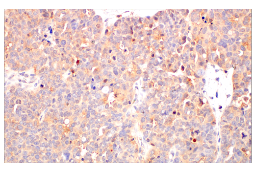
Immunohistochemical analysis of paraffin-embedded human ovarian serous adenocarcinoma using c-Rel (E8Z5Y) XP® Rabbit mAb.
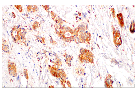
Immunohistochemical analysis of paraffin-embedded human ductal breast carcinoma using c-Rel (E8Z5Y) XP® Rabbit mAb.
Immunohistochemical analysis of paraffin-embedded human urothelial carcinoma using c-Rel (E8Z5Y) XP® Rabbit mAb.
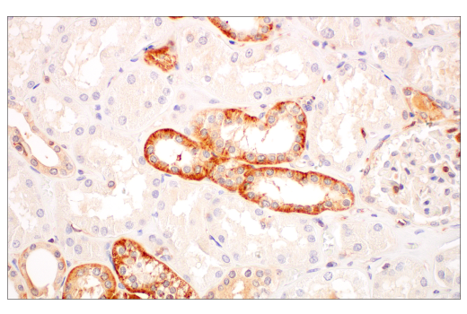
Immunohistochemical analysis of paraffin-embedded normal human kidney using c-Rel (E8Z5Y) XP® Rabbit mAb.
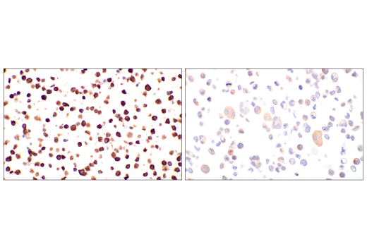
Immunohistochemical analysis of paraffin-embedded Ri-1 cell pellet (left, high-expressing) or MDA-MB-453 cell pellet (right, low-expressing) using c-Rel (E8Z5Y) XP® Rabbit mAb.
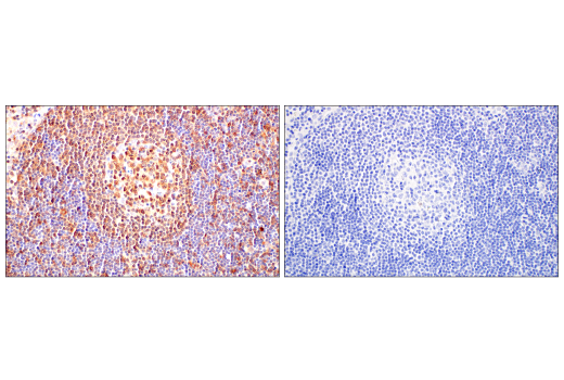
Immunohistochemical analysis of paraffin-embedded normal human lymph node using c-Rel (E8Z5Y) XP® Rabbit mAb (left) compared to concentration-matched Rabbit (DA1E) mAb IgG XP® Isotype Control #3900 (right).
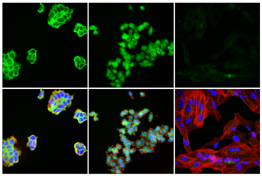
Confocal immunofluorescent analysis of RT-4 cells (high-expressing), either untreated (left) or treated with hTNF-α (20 ng/mL, 30 min; middle), and Caki-1 cells (right, low-expressing) using c-Rel (E8Z5Y) XP® Rabbit mAb (green), DyLight 650 Phalloidin #12956 (red), and DAPI #4083 (blue).