


 全部商品分类
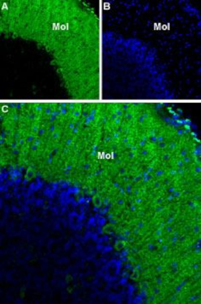


CACNA1G (CAV3.1) ANTIBODY

 下载产品说明书 下载SDS
 用小程序,查商品更便捷


 收藏
收藏
 对比
对比 咨询
咨询种属反应
宿主/亚型
分类
类型
抗原
偶联物
形式
浓度
纯化类型
保存液
内含物
保存条件
运输条件
产品详细信息
Reconstitution: 25 µL, 50 µL or 0.2 mL double distilled water (DDW), depending on the sample size. The antibody ships as a lyophilized powder at room temperature. Upon arrival, it should be stored at -20C. The reconstituted solution can be stored at 4C for up to 1 week. For longer periods, small aliquots should be stored at -20C. Avoid multiple freezing and thawing. Centrifuge all antibody preparations before use (10000 x g 5 min).
靶标信息
Voltage-dependent Ca2+ channels mediate Ca2+ entry into excitable cells in response to membrane depolarization, and they are involved in a variety of Ca2+-dependent processes, including muscle contraction, hormone or neurotransmitter release and gene expression. Calcium channels are highly diverse, multimeric complexes composed of an alpha-1 subunit, an intracellular beta subunit, a disulfide linked alpha-2/delta subunit and a transmembrane gamma subunit. Ca2+ currents are characterized on the basis of their biophysical and pharmacologic properties and include L-, N-, T-, P-, Q-, and R- types. T-type Ca2+ currents are activated and inactivated more rapidly and at more negative membrane potentials than other Ca2+ current types. T-type Ca2+ channels enhance odor sensitivity by lowering the threshold of spike generation in olfactory receptor cells (ORCs).
仅用于科研。不用于诊断过程。未经明确授权不得转售。
生物信息学
蛋白别名: calcium channel, voltage-dependent, T type, alpha 1G subunit; CaV T1; Cav3 1; cav3 1c; Cav3.1c; MGC117234; NBR13; voltage-dependent calcium channel alpha 1G subunit, isoform 11; voltage-dependent T-type calcium channel alpha 1G subunit; Voltage-dependent T-type calcium channel subunit alpha-1G; Voltage-gated calcium channel subunit alpha Cav3.1
基因别名: [a]1G; a1G; alpha-1G; Ca(V)T.1; CACNA1G; Cav3.1; Cav3.1d; KIAA1123; mKIAA1123; NBR13; SCA42
UniProt ID:(Human) O43497
Entrez Gene ID:(Human) 8913, (Rat) 29717, (Mouse) 12291
参考图片
Expression of CACNA1Gin rat cerebellum - Immunohistochemical staining of rat cerebellum using Anti-CACNA1G (CaV3.1) Antibody (#ACC-021). A. CACNA1Gimmunoreactivity (green) appears in the molecular layer. B. Nuclear staining using DAPI as the counterstain (blue). C. Merged images A and B. Mol = molecular layer.
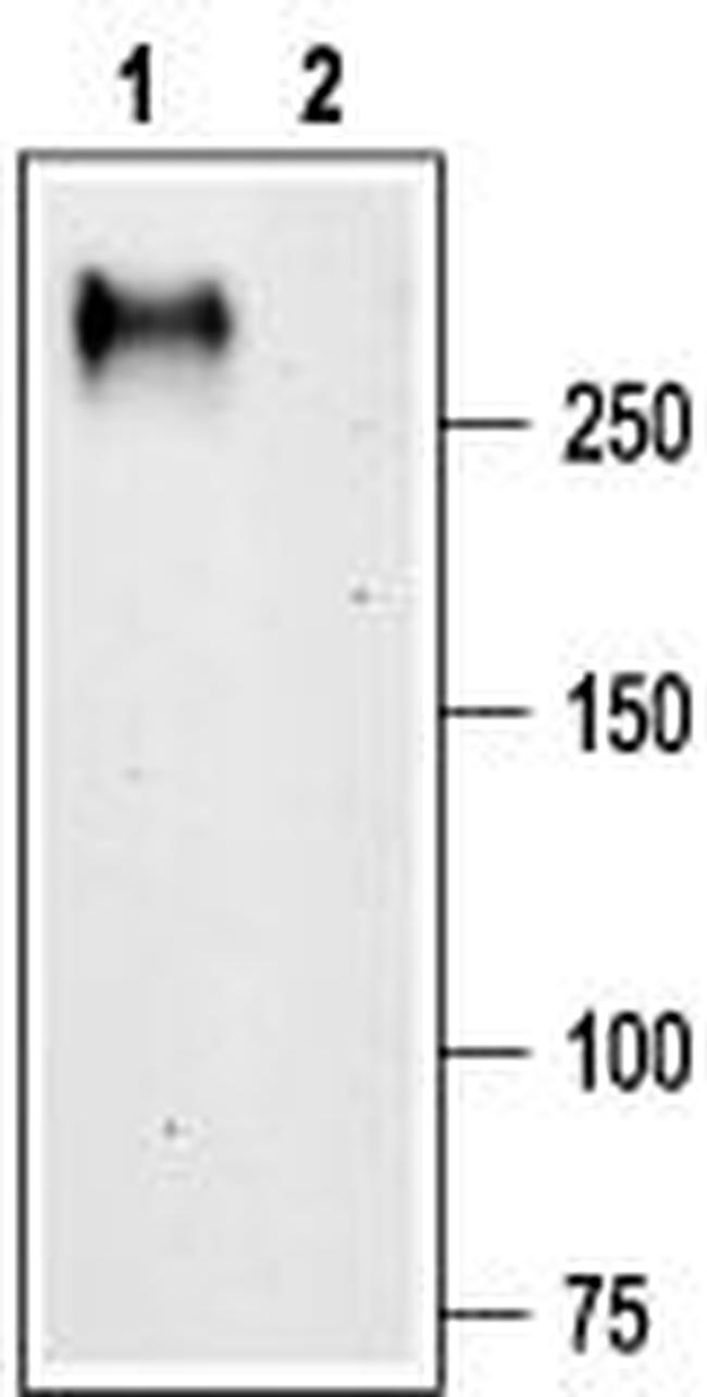
Western blot analysis of rat brain membranes: - 1. Anti-CACNA1G (CaV3.1) Antibody (#ACC-021), (1:200).2. Anti-CACNA1G (CaV3.1) Antibody , preincubated with CACNA1G/Cav3.1 Blocking Peptide (#BLP-CC021).