


 全部商品分类


N-Cadherin (D4R1H) XP ® Rabbit mAb


 下载产品说明书 下载COA 下载SDS
 用小程序,查商品更便捷


 收藏
收藏
 对比
对比 咨询
咨询
Monoclonal antibody is produced by immunizing animals with a synthetic peptide corresponding to residues surrounding Arg526 of human N-cadherin protein.
Product Usage Information
| Application | Dilution |
|---|---|
| Western Blotting | 1:1000 |
| Simple Western™ | 1:10 - 1:50 |
| Immunoprecipitation | 1:50 |
| IHC Leica Bond | 1:25 - 1:100 |
| Immunohistochemistry (Paraffin) | 1:50 - 1:200 |
| Immunofluorescence (Frozen) | 1:400 |
| Immunofluorescence (Immunocytochemistry) | 1:400 - 1:1600 |
Specificity/Sensitivity
Species Reactivity:
Human, Mouse
Supplied in 10 mM sodium HEPES (pH 7.5), 150 mM NaCl, 100 µg/ml BSA, 50% glycerol and less than 0.02% sodium azide. Store at –20°C. Do not aliquot the antibody.
For a carrier free (BSA and azide free) version of this product see product #84117.
参考图片
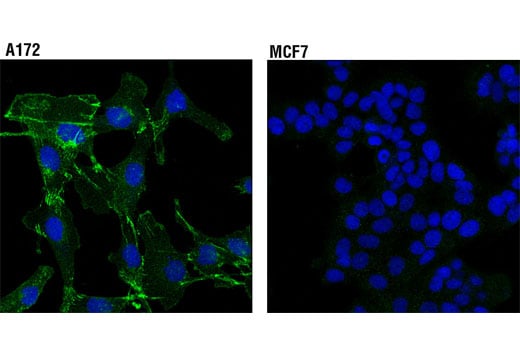
Confocal immunofluorescent analysis of A172 (positive, left) and MCF7 (negative, right) cells using N-Cadherin (D4R1H) XP® Rabbit mAb (green). Blue pseudocolor= DRAQ5® #4084 (fluorescent DNA dye).
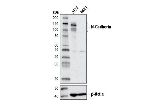
Western blot analysis of extracts from A172 and MCF7 cells using N-Cadherin (D4R1H) XP® Rabbit mAb (upper) or β-Actin (D6A8) Rabbit mAb #8457 (lower).
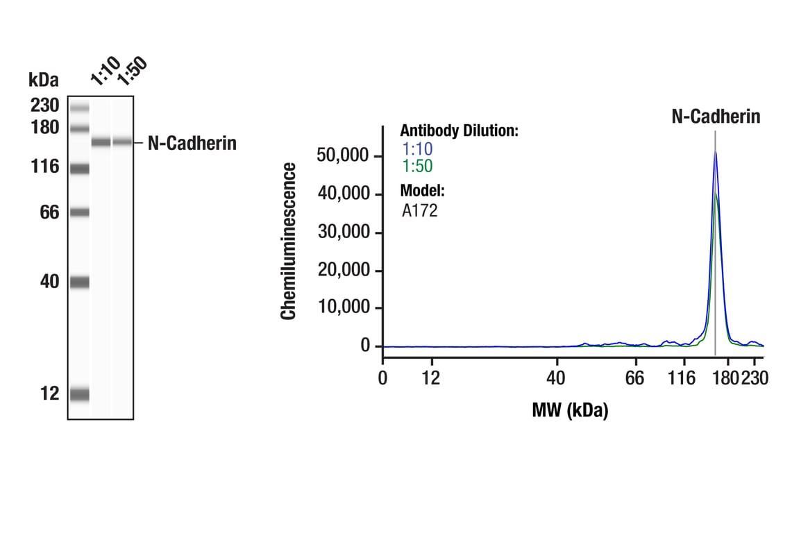
Simple Western™ analysis of lysates (0.1 mg/mL) from A172 lysate using N-Cadherin (D4R1H) XP® Rabbit mAb #13116. The virtual lane view (left) shows the target band (as indicated) at 1:10 and 1:50 dilutions of primary antibody. The corresponding electropherogram view (right) plots chemiluminescence by molecular weight along the capillary at 1:10 (blue line) and 1:50 (green line) dilutions of primary antibody. This experiment was performed under reducing conditions on the Jess™ Simple Western instrument from ProteinSimple, a BioTechne brand, using the 12-230 kDa separation module.
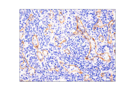
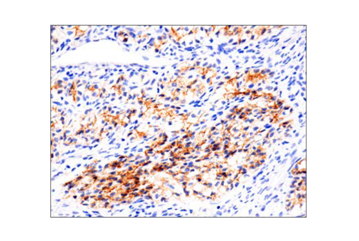
Immunohistochemical analysis of paraffin-embedded human non-Hodgkin Lymphoma using N-Cadherin (D4R1H) XP® Rabbit mAb performed on the Leica® BOND™ Rx.
Immunohistochemical analysis of paraffin-embedded human granulosa cell tumor of the ovary using N-Cadherin (D4R1H) XP® Rabbit mAb performed on the Leica® BOND™ Rx.
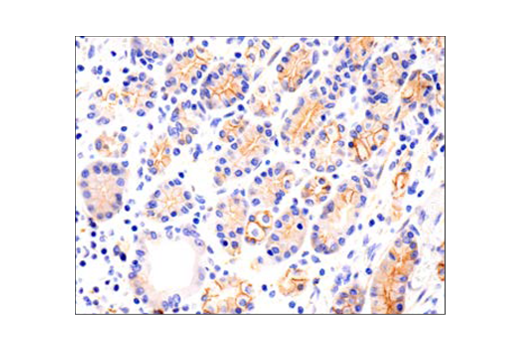
Immunohistochemical analysis of paraffin-embedded human gastric carcinoma using N-Cadherin (D4R1H) XP® Rabbit mAb performed on the Leica® BOND™ Rx.
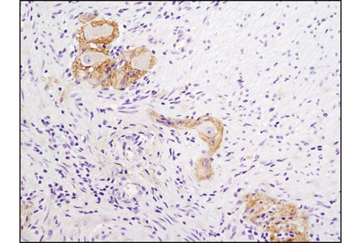
Immunohistochemical analysis of paraffin-embedded human colon using N-Cadherin (D4R1H) XP® Rabbit mAb. Note staining of myenteric plexus.
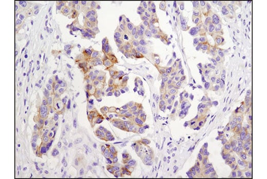
Immunohistochemical analysis of paraffin-embedded human ovarian carcinoma using N-Cadherin (D4R1H) XP® Rabbit mAb.
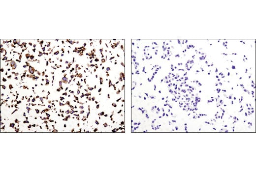
Immunohistochemical analysis of paraffin-embedded A172 (positive, left) and MCF7 (negative, right) cell pellets using N-Cadherin (D4R1H) XP® Rabbit mAb.
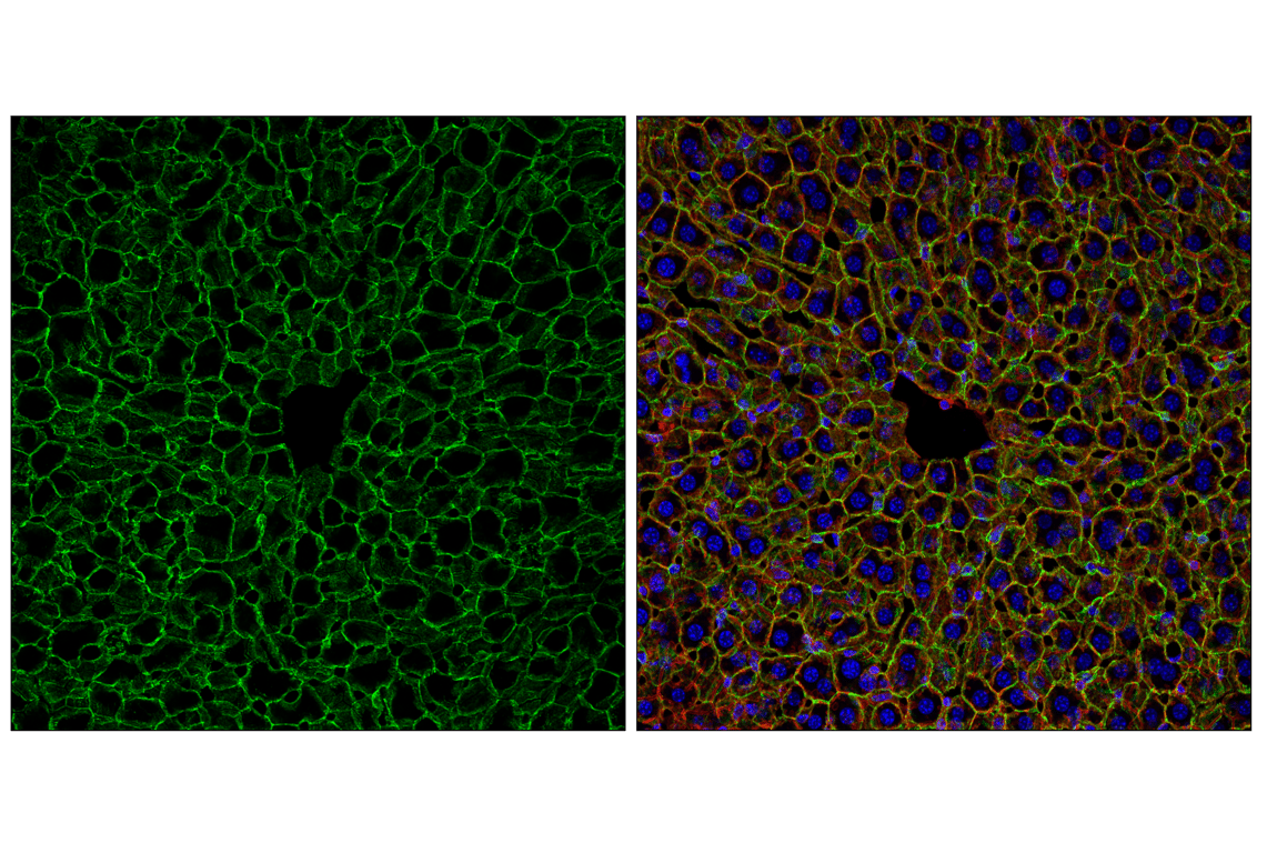
Confocal immunofluorescent analysis of fixed frozen mouse liver labeled with N-Cadherin (D4R1H) XP® Rabbit mAb (green), DyLight 554 Phalloidin #13054 (red) and ProLong® Gold Antifade Reagent with DAPI #8961 (blue).
Confocal immunofluorescent analysis of fixed frozen mouse choroid plexus labeled with N-Cadherin (D4R1H) XP® Rabbit mAb (green) and ProLong® Gold Antifade Reagent with DAPI #8961 (blue).
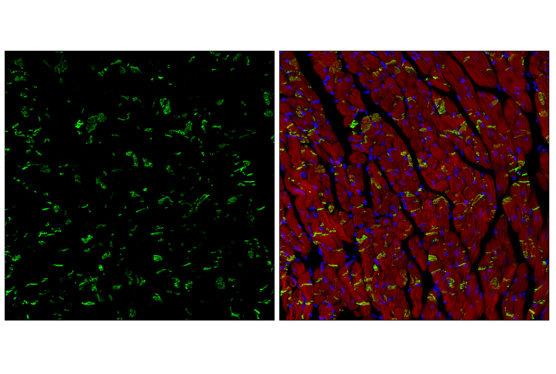
Confocal immunofluorescent analysis of fixed frozen mouse heart labeled with N-Cadherin (D4R1H) XP® Rabbit mAb (green), DyLight 554 Phalloidin #13054 (red) and ProLong® Gold Antifade Reagent with DAPI #8961 (blue).