


 全部商品分类
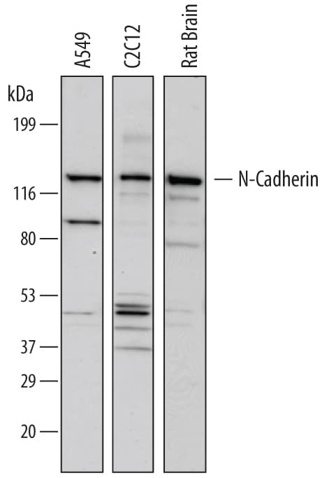


 下载产品说明书 下载SDS
 用小程序,查商品更便捷


 收藏
收藏
 对比
对比 咨询
咨询Flow Cytometry(2.5 µg/106 cells)
Immunohistochemistry(8-25 µg/mL)

Asp160-Ala724
Accession # P19022.4
Scientific Data
 View Larger
Detection of Human, Mouse, and Rat N‑Cadherin by Western Blot. Western blot shows lysates of A549 human lung carcinoma cell line, C2C12 mouse myoblast cell line, and rat brain tissue. PVDF membrane was probed with 1 µg/mL of Mouse Anti-Human N-Cadherin Monoclonal Antibody (Catalog # MAB13881) followed by HRP-conjugated Anti-Mouse IgG Secondary Antibody (Catalog # HAF007). A specific band was detected for N-Cadherin at approximately 130 kDa (as indicated). This experiment was conducted under reducing conditions and using Immunoblot Buffer Group 1.
 View Larger
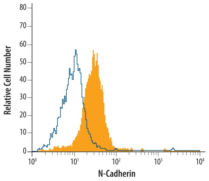
Detection of N-Cadherin in HeLa Human Cell Line by Flow Cytometry. HeLa human cervical epithelial carcinoma cell line was stained with Mouse Anti-Human N-Cadherin Monoclonal Antibody (Catalog # MAB13881, filled histogram) or isotype control antibody mouse IgM (open histogram), followed by Phycoerythrin-conjugated Anti-Mouse IgM Secondary Antibody (Catalog # F0116).
.") View Larger
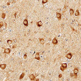
View LargerN‑Cadherin in Human Brain. N-Cadherin was detected in immersion fixed paraffin-embedded sections of Alzheimer's human brain using Mouse Anti-Human N-Cadherin Monoclonal Antibody (Catalog # MAB13881) at 15 µg/mL overnight at 4 °C. Before incubation with the primary antibody, tissue was subjected to heat-induced epitope retrieval using Antigen Retrieval Reagent-Basic (Catalog # CTS013). Tissue was stained using the Anti-Mouse HRP-DAB Cell & Tissue Staining Kit (brown; Catalog # CTS002) and counterstained with hematoxylin (blue). Specific staining was localized to cytoplasm of neurons. View our protocol for Chromogenic IHC Staining of Paraffin-embedded Tissue Sections.
Human N-Cadherin Antibody Summary
Asp160-Ala724
Accession # P19022.4
Applications
Please Note: Optimal dilutions should be determined by each laboratory for each application. General Protocols are available in the Technical Information section on our website.
Flow Cytometry(2.5 µg/106 cells)
Immunohistochemistry(8-25 µg/mL)
Background: N-Cadherin
Neuronal Cadherin (N-Cadherin or NCAD), also known as Cadherin-2 (CDH2), is a 130 kDa type I membrane protein belonging to the Cadherin superfamily of calcium-dependent adhesion molecules. Cadherins are involved in multiple processes including embryonic development, cell migration, and maintenance of epithelial integrity (1, 2). Human N‑Cadherin is synthesized with a 25 amino acid (aa) signal peptide and a 134 aa N‑terminal propeptide. The mature cell surface‑expressed protein consists of a 565 amino acid (aa) extracellular domain (ECD) that contains five Cadherin repeats, a 21 aa transmembrane segment, and a 161 aa cytoplasmic domain (3). Within the ECD, human N-Cadherin shares 98% aa sequence identity with mouse and rat N-Cadherin. In the nervous system, N-Cadherin mediates adhesion between the opposing faces of developing neuronal synapses and between Schwann cells and neuronal axons (4, 5). It interacts in cis or in trans homophilically and with the GluR2 subunit of neuronal AMPA receptors (1, 6). During synaptic maturation, its expression is lost from inhibitory terminals but maintained at excitatory terminals (5). ADAM10-mediated shedding of the N-Cadherin ECD alters cell-cell adhesion, synaptic development, and AMPA receptor activity (7, 8). N-Cadherin can also be cleaved at multiple additional sites within the intracellular or extracellular domains by Calpain, gamma ‑Secretase, and several MMPs (9 - 13). Cleavage of N‑Cadherin in atherosclerotic plaques contributes alternatively to vascular smooth muscle cell proliferation (MMP-9 and -12) or apoptosis (MMP‑7) (12, 13). Aberrant cell surface expression of the pro and mature forms of N-Cadherin in cancer results in increased tumor progression and invasiveness (14, 15). N-Cadherin also mediates the adhesion between hematopoeitic progenitor cells and mesenchymal stromal cells of the bone marrow (16).
- Pokutta, S. and W.I. Weis (2007) Annu. Rev. Cell Dev. Biol. 23:237.
- Gumbiner, B.M. (2005) Nat. Rev. Mol. Cell Biol. 6:622.
- Reid, R.A. and J.J. Hemperly (1990) Nucleic Acids Res. 18:5896.
- Wanner, I.B. and P.M. Wood (2002) J. Neurosci. 22:4066.
- Benson, D.L. and H. Tanaka (1998) J. Neurosci. 18:6892.
- Saglietti, L. et al. (2007) Neuron 54:461.
- Reiss, K. et al. (2005) EMBO J. 24:742.
- Malinverno, M. et al. (2010) J. Neurosci. 30:16343.
- Jang, Y.-N. et al. (2009) J. Neurosci. 29:5974.
- Uemura, K. et al. (2006) Neurosci. Lett. 402:278.
- Hartland, S.N. et al. (2009) Liver Int. 29:966.
- Williams, H. et al. (2010) Cardiovasc. Res. 87:137.
- Dwivedi, A. et al. (2009) Cardiovasc. Res. 81:178.
- Maret, D. et al. (2010) Neoplasia 12:1066.
- Tanaka, H. et al. (2010) Nat. Med. 16:1414.
- Wein, F. et al. (2010) Stem Cell Res. 4:129.
Preparation and Storage
- 12 months from date of receipt, -20 to -70 °C as supplied.
- 1 month, 2 to 8 °C under sterile conditions after reconstitution.
- 6 months, -20 to -70 °C under sterile conditions after reconstitution.
参考图片
Detection of Human, Mouse, and Rat N‑Cadherin by Western Blot. Western blot shows lysates of A549 human lung carcinoma cell line, C2C12 mouse myoblast cell line, and rat brain tissue. PVDF membrane was probed with 1 µg/mL of Mouse Anti-Human N‑Cadherin Monoclonal Antibody (Catalog # MAB13881) followed by HRP-conjugated Anti-Mouse IgG Secondary Antibody (Catalog # HAF007). A specific band was detected for N‑Cadherin at approximately 130 kDa (as indicated). This experiment was conducted under reducing conditions and using Immunoblot Buffer Group 1.
Detection of N-Cadherin in HeLa Human Cell Line by Flow Cytometry. HeLa human cervical epithelial carcinoma cell line was stained with Mouse Anti-Human N‑Cadherin Monoclonal Antibody (Catalog # MAB13881, filled histogram) or isotype control antibody mouse IgM (open histogram), followed by Phycoerythrin-conjugated Anti-Mouse IgM Secondary Antibody (Catalog # F0116).
N‑Cadherin in Human Brain. N‑Cadherin was detected in immersion fixed paraffin-embedded sections of Alzheimer's human brain using Mouse Anti-Human N‑Cadherin Monoclonal Antibody (Catalog # MAB13881) at 15 µg/mL overnight at 4 °C. Before incubation with the primary antibody, tissue was subjected to heat-induced epitope retrieval using Antigen Retrieval Reagent-Basic (Catalog # CTS013). Tissue was stained using the Anti-Mouse HRP-DAB Cell & Tissue Staining Kit (brown; Catalog # CTS002) and counterstained with hematoxylin (blue). Specific staining was localized to cytoplasm of neurons. View our protocol for Chromogenic IHC Staining of Paraffin-embedded Tissue Sections.