


 全部商品分类
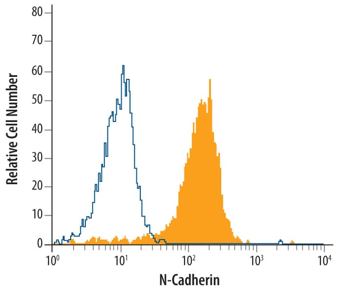


 下载产品说明书 下载SDS
 用小程序,查商品更便捷


 收藏
收藏
 对比
对比 咨询
咨询
Ser26-Arg159
Accession # P19022
Scientific Data
 View Larger
Detection of N-Cadherin in HeLa Human Cell Line by Flow Cytometry. HeLa human cervical epithelial carcinoma cell line was stained with Mouse Anti-Human N-Cadherin Monoclonal Antibody (Catalog # MAB13882, filled histogram) or isotype control antibody mouse IgM (open histogram), followed by Phycoerythrin-conjugated Anti-Mouse IgM Secondary Antibody (Catalog # F0116).
Human N-Cadherin Antibody Summary
Ser26-Arg159
Accession # P19022
Applications
Please Note: Optimal dilutions should be determined by each laboratory for each application. General Protocols are available in the Technical Information section on our website.
Background: N-Cadherin
N‑Cadherin (Neural Cadherin; also CD325 and Cadherin-2) is a 130‑135 kDa member of the "classical" (or type I) cadherin subfamily, cadherin superfamily of proteins. It is expressed on multiple cell types, including neurons, fibroblasts, Schwann cells, endothelial cells and hepatic stellate cells. N‑Cadherin mediates homotypic binding, either in cis (same cell) or trans (adjacent cell). proN‑Cadherin is expressed as an 881 amino acid (aa) type I transmembrane glycoprotein. It may be initially inserted into the ER, where the 15‑20 kDa prodomain (aa 26‑159) is cleaved by proprotein convertase, and the mature molecule is transported to the surface. Alternatively, on neurons, proN‑Cadherin may first appear on the surface, with cleavage occurring at the time of synaptogenesis. Cleavage appears necessary for homophilic interaction as presence of the prodomain is suggested to negatively regulate oligomer formation. Over the entire prodomain, the human N‑Cadherin proregion shares 87% aa identity with the mouse N‑Cadherin proregion.
Preparation and Storage
- 12 months from date of receipt, -20 to -70 °C as supplied.
- 1 month, 2 to 8 °C under sterile conditions after reconstitution.
- 6 months, -20 to -70 °C under sterile conditions after reconstitution.
参考图片
Detection of N-Cadherin in HeLa Human Cell Line by Flow Cytometry. HeLa human cervical epithelial carcinoma cell line was stained with Mouse Anti-Human N‑Cadherin Monoclonal Antibody (Catalog # MAB13882, filled histogram) or isotype control antibody mouse IgM (open histogram), followed by Phycoerythrin-conjugated Anti-Mouse IgM Secondary Antibody (Catalog # F0116).