


 全部商品分类


Caspase-3 (D3R6Y) Rabbit mAb


 下载产品说明书 下载COA 下载SDS
 用小程序,查商品更便捷


 收藏
收藏
 对比
对比 咨询
咨询
Monoclonal antibody is produced by immunizing animals with recombinant protein specific to the p20 subunit of human caspase-3 protein.
Product Usage Information
| Application | Dilution |
|---|---|
| Western Blotting | 1:1000 |
| Simple Western™ | 1:10 - 1:50 |
| Immunoprecipitation | 1:100 |
Specificity/Sensitivity
Species Reactivity:
Human, Mouse, Rat, Monkey
Supplied in 10 mM sodium HEPES (pH 7.5), 150 mM NaCl, 100 µg/ml BSA, 50% glycerol and less than 0.02% sodium azide. Store at –20°C. Do not aliquot the antibody.
参考图片
Simple Western™ analysis of lysates (1 mg/mL) from Jurkat cells treated with Cytochrome C using Caspase-3 (D3R6Y) Rabbit mAb #14220. The virtual lane view (left) shows the target bands (as indicated) at 1:10 and 1:50 dilutions of primary antibody. The corresponding electropherogram view (right) plots chemiluminescence by molecular weight along the capillary at 1:10 (blue line) and 1:50 (green line) dilutions of primary antibody. This experiment was performed under reducing conditions on the Jess™ Simple Western instrument from ProteinSimple, a BioTechne brand, using the 12-230 kDa separation module.
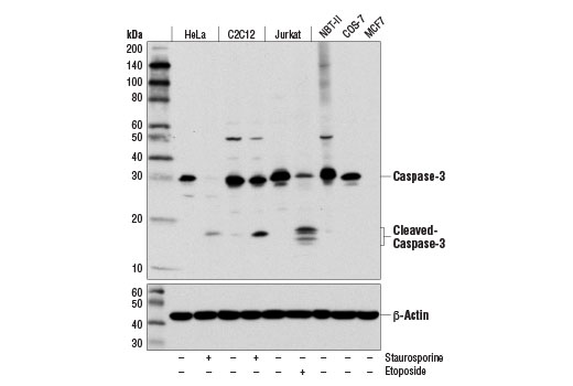
Western blot analysis of various cell lines, untreated (-) or treated with Staurosporine #9953 (1 μM; 3 hr) or with Etoposide #2200 (25 μM, overnight), using Caspase-3 (D3R6Y) Rabbit mAb (upper) or β-Actin (D6A8) Rabbit mAb #8457 (lower). MCF7 cells are negative for caspase-3 expression.
Western blot analysis of extracts from HCT116 cells (lane 1) or CASP3 knock-out cells (lane 2) using Caspase-3 (D3R6Y) Rabbit mAb #14220 (upper), and α-Actinin (D6F6) XP® Rabbit mAb #6487 (lower). The absence of signal in the CASP3 knock-out HCT116 cells confirms specificity of the antibody for CASP3.