


 全部商品分类
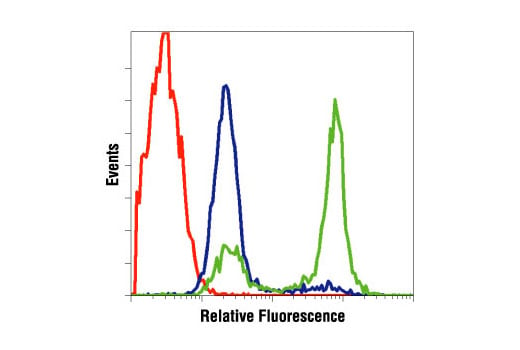


Cleaved Caspase-3 (Asp175) Antibody

 下载产品说明书 下载COA 下载SDS
 用小程序,查商品更便捷


 收藏
收藏
 对比
对比 咨询
咨询
Polyclonal antibodies are produced by immunizing animals with a synthetic peptide corresponding to amino-terminal residues adjacent to (Asp175) in human caspase-3.
Product Usage Information
| Application | Dilution |
|---|---|
| Western Blotting | 1:1000 |
| Simple Western™ | 1:10 - 1:50 |
| Immunoprecipitation | 1:100 |
| Immunohistochemistry (Paraffin) | 1:400 |
| Immunofluorescence (Immunocytochemistry) | 1:400 |
| Flow Cytometry (Fixed/Permeabilized) | 1:800 |
Specificity/Sensitivity
Species Reactivity:
Human, Mouse, Rat, Monkey
Supplied in 10 mM sodium HEPES (pH 7.5), 150 mM NaCl, 100 µg/ml BSA and 50% glycerol. Store at –20°C. Do not aliquot the antibody.
参考图片
Flow cytometric analysis of Jurkat cells, untreated (blue) or treated with etoposide #2200 (green), using Cleaved Caspase-3 (Asp175) Antibody compared to a nonspecific negative control antibody (red).
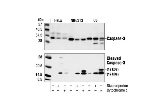
Western blot analysis of extracts from HeLa, NIH/3T3 and C6 cells untreated, staurosporine-treated (3hrs, 1 µM in vivo) or cytochrome c-treated (1hr, 0.25 mg/ml in vitro), using Caspase-3 Antibody #9662 (upper) or Cleaved Caspase-3 (Asp175) Antibody (lower).
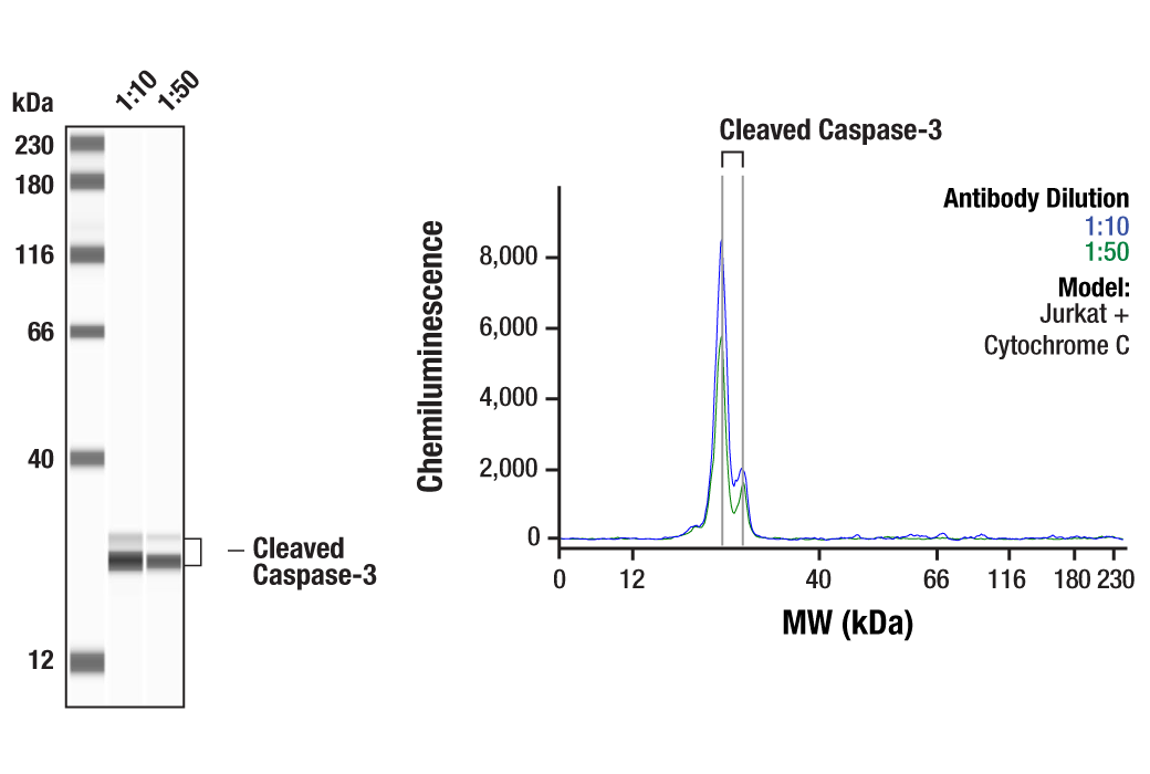
Simple Western™ analysis of lysates (0.1 mg/mL) from Jurkat cells treated with Cytochrome C using Cleaved Caspase-3 (Asp175) Antibody #9661. The virtual lane view (left) shows two target bands (as indicated) at 1:10 and 1:50 dilutions of primary antibody. The corresponding electropherogram view (right) plots chemiluminescence by molecular weight along the capillary at 1:10 (blue line) and 1:50 (green line) dilutions of primary antibody. This experiment was performed under reducing conditions on the Jess™ Simple Western instrument from ProteinSimple, a BioTechne brand, using the 12-230 kDa separation module.
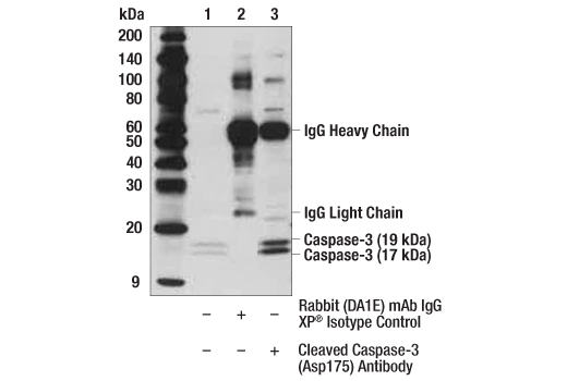
Immunoprecipitation of cleaved caspase-3 from Jurkat extracts treated with Etoposide #2200 (25 mM; 5 hr). Lane 1 is 10% input, lane 2 is Rabbit (DA1E) mAb IgG XP® Isotype Control #3900, and lane 3 is Cleaved Caspase-3 (Asp175) Antibody. Western blot analysis was performed using Cleaved Caspase-3 (Asp175) Antibody. Anti-rabbit IgG, HRP-linked Antibody #7074 was used as a secondary antibody.
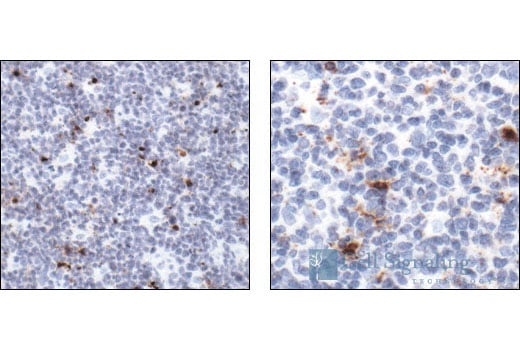
Immunohistochemical analysis of paraffin-embedded human tonsil, showing cytoplasmic and perinuclear localization in apoptotic cells (low and high magnification), using Cleaved Caspase-3 (Asp175) Antibody.
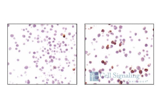
Immunohistochemical analysis using Cleaved caspase-3 (Asp175) antibody on SignalSlide™ Cleaved Caspase-3 IHC controls #8104 (paraffin-embedded Jurkat cells, untreated (left) or etoposide-treated (right)).
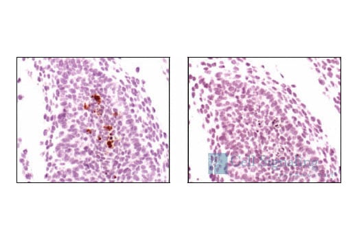
Immunohistochemical analysis of paraffin-embedded mouse embryo, using Cleaved Caspase-3 (Asp175) Antibody preincubated with control peptide (left) or Cleaved Caspase-3 (Asp175) Blocking Peptide #1050 (right).
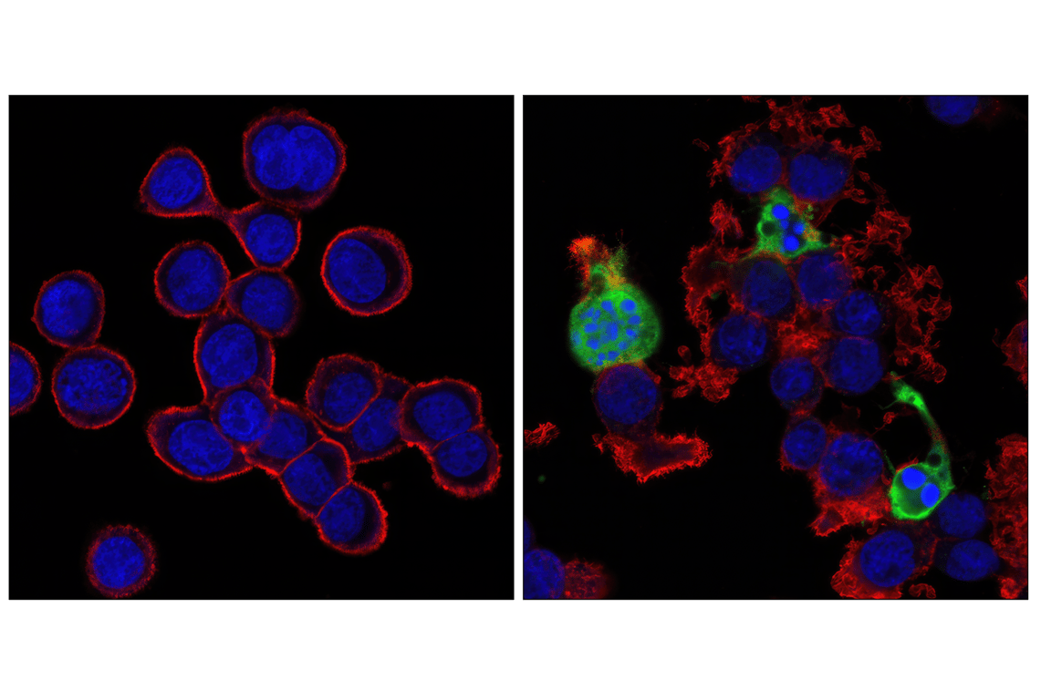
Confocal immunofluorescent images of HT-29 cells, untreated (left) or Staurosporine #9953 treated (right), labeled with Cleaved Caspase-3 (Asp175) Antibody (green). Actin filaments have been labeled with Alexa Fluor® 555 phalloidin #8953 (red). Blue pseudocolor = DRAQ5® (fluorescent DNA dye).