


 全部商品分类
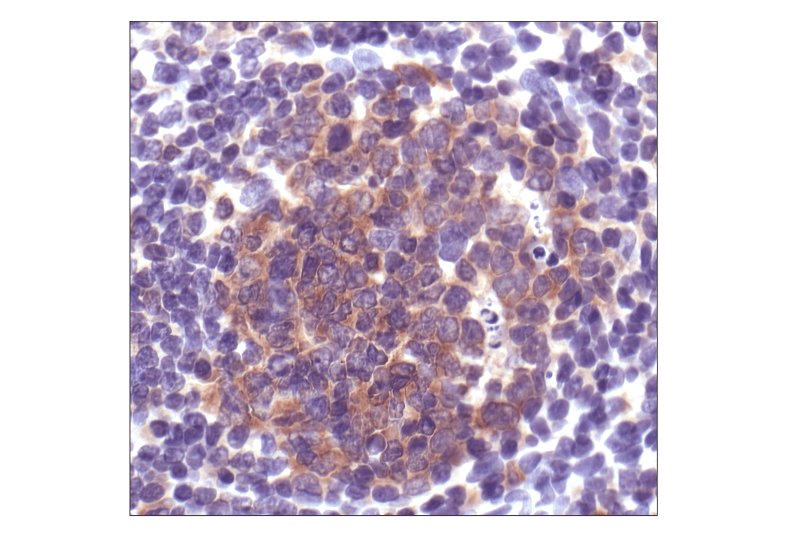


Caspase-3 Antibody


 下载产品说明书 下载COA 下载SDS
 用小程序,查商品更便捷


 收藏
收藏
 对比
对比 咨询
咨询
Polyclonal antibodies are produced by immunizing animals with a synthetic peptide corresponding to residues surrounding the cleavage site of human caspase-3. Antibodies are purified by protein A and peptide affinity chromatography.
Product Usage Information
| Application | Dilution |
|---|---|
| Western Blotting | 1:1000 |
| Simple Western™ | 1:10 - 1:50 |
| Immunoprecipitation | 1:50 |
| Immunohistochemistry (Paraffin) | 1:1000 |
Specificity/Sensitivity
Species Reactivity:
Human, Mouse, Rat, Monkey
Supplied in 10 mM sodium HEPES (pH 7.5), 150 mM NaCl, 100 µg/ml BSA and 50% glycerol. Store at –20°C. Do not aliquot the antibody.
参考图片
Immunohistochemical staining of paraffin-embedded human tonsil, showing cytoplasmic localization, using Caspase-3 Antibody.
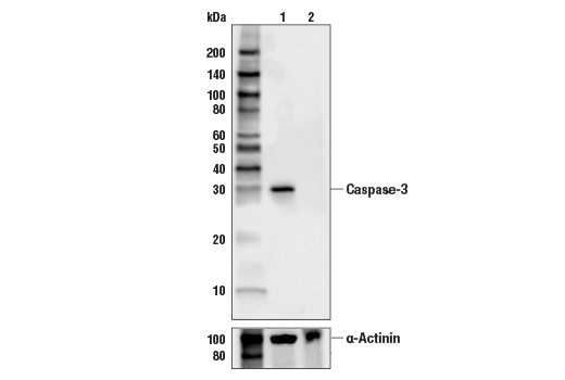
Western blot analysis of extracts from HCT116 cells (lane 1) or CASP3 knock-out cells (lane 2) using Caspase-3 Antibody #9662 (upper), and α-Actinin (D6F6) XP® Rabbit mAb #6487 (lower). The absence of signal in the CASP3 knock-out HCT116 cells confirms specificity of the antibody for CASP3.
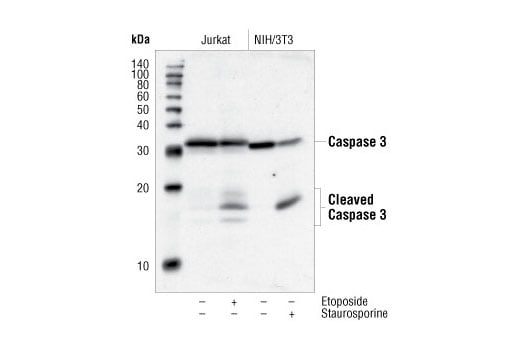
Western blot analysis of extracts from Jurkat cells, untreated or etoposide-treated (25uM, 5hrs), and NIH/3T3 cells, untreated or staurosporine-treated (1uM, 3hrs), using Caspase-3 Antibody.
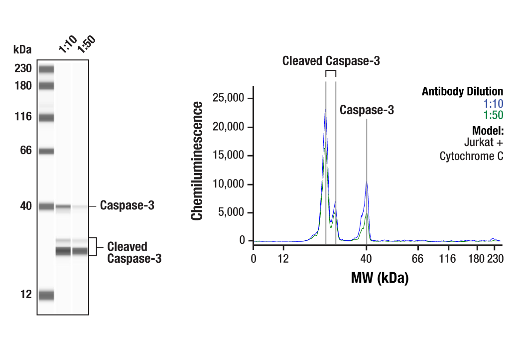
Simple Western™ analysis of lysates (0.1 mg/mL) from Jurkat cells treated with Cytochrome C using Caspase-3 Antibody #9662. The virtual lane view (left) shows the target bands (as indicated) at 1:10 and 1:50 dilutions of primary antibody. The corresponding electropherogram view (right) plots chemiluminescence by molecular weight along the capillary at 1:10 (blue line) and 1:50 (green line) dilutions of primary antibody. This experiment was performed under reducing conditions on the Jess™ Simple Western instrument from ProteinSimple, a BioTechne brand, using the 12-230 kDa separation module.