


 全部商品分类
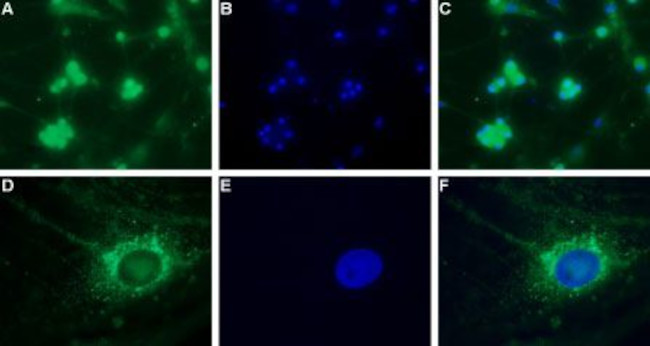


CAV3.2 (CACNA1H) ANTIBODY

 下载产品说明书 下载SDS
 用小程序,查商品更便捷


 收藏
收藏
 对比
对比 咨询
咨询种属反应
宿主/亚型
分类
类型
抗原
偶联物
形式
浓度
纯化类型
保存液
内含物
保存条件
运输条件
产品详细信息
Reconstitution: 25 µL, 50 µL or 0.2 mL double distilled water (DDW), depending on the sample size. The antibody ships as a lyophilized powder at room temperature. Upon arrival, it should be stored at -20C. The reconstituted solution can be stored at 4C for up to 1 week. For longer periods, small aliquots should be stored at -20C. Avoid multiple freezing and thawing. Centrifuge all antibody preparations before use (10000 x g 5 min).
靶标信息
This gene encodes a T-type member of the alpha-1 subunit family, a protein in the voltage-dependent calcium channel complex. Calcium channels mediate the influx of calcium ions into the cell upon membrane polarization and consist of a complex of alpha-1, alpha-2/delta, beta, and gamma subunits in a 1:1:1:1 ratio. The alpha-1 subunit has 24 transmembrane segments and forms the pore through which ions pass into the cell. There are multiple isoforms of each of the proteins in the complex, either encoded by different genes or the result of alternative splicing of transcripts. Alternate transcriptional splice variants, encoding different isoforms, have been characterized for the gene described here. Studies suggest certain mutations in this gene lead to childhood absence epilepsy (CAE).
仅用于科研。不用于诊断过程。未经明确授权不得转售。
生物信息学
蛋白别名: A1H-b; calcium channel alpha13.2 subunit; calcium channel, voltage-dependent, T type, alpha 1H subunit; calcium channel, voltage-dependent, T type, alpha 1Hb subunit; FLJ90484; Low-voltage-activated calcium channel alpha1 3.2 subunit; low-voltage-activated calcium channel alpha13.2 subunit; T-type Cav3.2; voltage dependent t-type calcium channel alpha-1H subunit; Voltage-dependent T-type calcium channel subunit alpha-1H; voltage-gated calcium channel alpha subunit Cav3.2; voltage-gated calcium channel alpha subunit CavT.2; Voltage-gated calcium channel subunit alpha Cav3.2
基因别名: alpha13.2; CACNA1H; CACNA1HB; Cav3.2; ECA6; EIG6; MNCb-1209
UniProt ID:(Human) O95802, (Rat) Q9EQ60
Entrez Gene ID:(Human) 8912, (Rat) 114862, (Mouse) 58226
参考图片
Expression of CaV3.2 in rat DRG primary culture - Immunocytochemical staining of paraformaldehyde-fixed and permeabilized rat dorsal root ganglion (DRG) primary culture. A, D.Immunocytochemical staining using Anti-CaV3.2 (CACNA1H) Antibody (#ACC-025), (1:200), followed by goat Anti-rabbit-AlexaFluor-488 secondary Antibody . B, E. Nuclear fluorescence staining of cells using the membrane-permeable DNA dye Hoechst 33342.C. Merged image of panels A and B.F. Merged image of panels D and E.Magnification:A-C: x20D-F: x100.
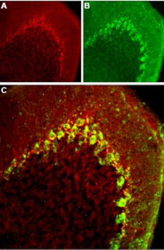
Expression of CaV3.2 in mouse cerebellum - Immunohistochemical staining of mouse cerebellum frozen sections with Anti-CaV3.2 (CACNA1H) Antibody (#ACC-025), (1:100). A. CaV3.2 appears adjacent to Purkinje cells and in fibers in the molecular layer (red). B. Staining of Purkinje cells with mouse Anti-parvalbumin (PV, green). C. Merged image of panels A and B demonstrates presence of CaV3.2 adjacent to Purkinjecells.
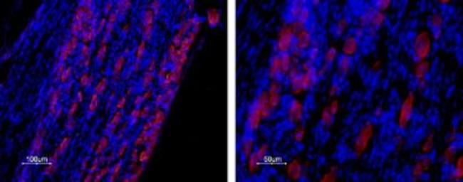
Expression of CaV3.2 in rat DRG - Immunohistochemical staining of rat dorsal root ganglion (DRG) frozen sections with Anti-CaV3.2 (CACNA1H) Antibody (#ACC-025), (1:50). Staining is specific for DRG. Note that neither glial cells nor axonal fibers are stained. Hoechst 33342 is used as the counterstain.
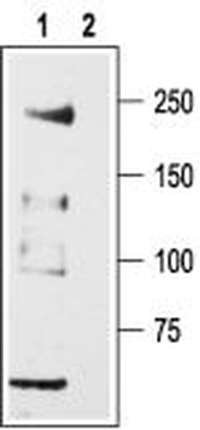
Western blot analysis of rat DRG lysates: - 1. Anti-CaV3.2 (CACNA1H) Antibody (#ACC-025), (1:200).2. Anti-CaV3.2 (CACNA1H) Antibody , preincubated with Cav3.2/CACNA1H Blocking Peptide (#BLP-CC025).