


 全部商品分类
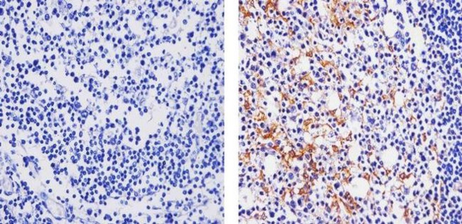


 下载产品说明书 下载COA 下载SDS
 用小程序,查商品更便捷


 收藏
收藏
 对比
对比 咨询
咨询种属反应
宿主/亚型
Expression System
分类
类型
克隆号
抗原
偶联物
形式
浓度
纯化类型
保存液
内含物
保存条件
运输条件
RRID
产品详细信息
Intact IgG appears on a non-reducing gel as ~150 kDa band and upon reduction generating a ~25 kDa light chain band and a ~50 kDa heavy chain.
Recombinant rabbit monoclonal antibodies are produced using in vitro expression systems. The expression systems are developed by cloning in the specific antibody DNA sequences from immunoreactive rabbits. Then, individual clones are screened to select the best candidates for production. The advantages of using recombinant rabbit monoclonal antibodies include: better specificity and sensitivity, lot-to-lot consistency, animal origin-free formulations, and broader immunoreactivity to diverse targets due to larger rabbit immune repertoire.
靶标信息
Both MIP-1alpha and MIP-1beta are structurally and functionally related CC chemokines. They participate in host response to invading bacterial, viral, parasite and fungal pathogens by regulating the trafficking and activation state of selected subgroups of inflammatory cells (e.g. macrophages, lymphocytes and NK cells). While both MIP-1alpha and MIP-1beta exert similar effects on monocytes, their effect on lymphocytes differ; with MIP-1alpha selectively attracting CD8+ lymphocytes, and MIP-1beta selectively attracting CD4+ lymphocytes. Additionally, MIP-1alpha and MIP-1beta have also been shown to be potent chemoattractants for B cells, eosinophils and dendritic cells. Both human and murine MIP-1alpha and MIP-1beta are active on human and murine hematopoietic cells.
仅用于科研。不用于诊断过程。未经明确授权不得转售。
生物信息学
蛋白别名: C-C motif chemokine 3; chemokine (C-C motif) ligand 3; G0/G1 switch regulatory protein 19-1; H-MIP-1-alpha; LD78-alpha; Macrophage inflammatory protein 1-alpha; MIP-1-alpha; mip1 alpha; PAT 464.1; RP23-320E6.7; SIS-beta; small inducible cytokine A3 (homologous to mouse Mip-1a); Small-inducible cytokine A3; Tonsillar lymphocyte LD78 alpha protein
基因别名: CCL3; G0S19-1; LD78ALPHA; MIP-1-alpha; MIP1A; SCYA3
UniProt ID:(Human) P10147
Entrez Gene ID:(Human) 6348
参考图片
Immunohistochemistry analysis of MIP1A showing staining in the cytoplasm of paraffin-embedded human tonsil tissue (right) compared to a negative control without primary antibody (left). To expose target proteins, antigen retrieval was performed using 10mM sodium citrate (pH 6.0), microwaved for 8-15 min. Following antigen retrieval, tissues were blocked in 3% H2O2-methanol for 15 min at room temperature, washed with ddH2O and PBS, and then probed with a MIP1A Recombinant Rabbit Monoclonal Antibody (Product # 701097) diluted in 3% BSA-PBS at a dilution of 1:20 for 1 hour at 37ºC in a humidified chamber. Tissues were washed extensively in PBST and detection was performed using an HRP-conjugated secondary antibody followed by colorimetric detection using a DAB kit. Tissues were counterstained with hematoxylin and dehydrated with ethanol and xylene to prep for mounting.
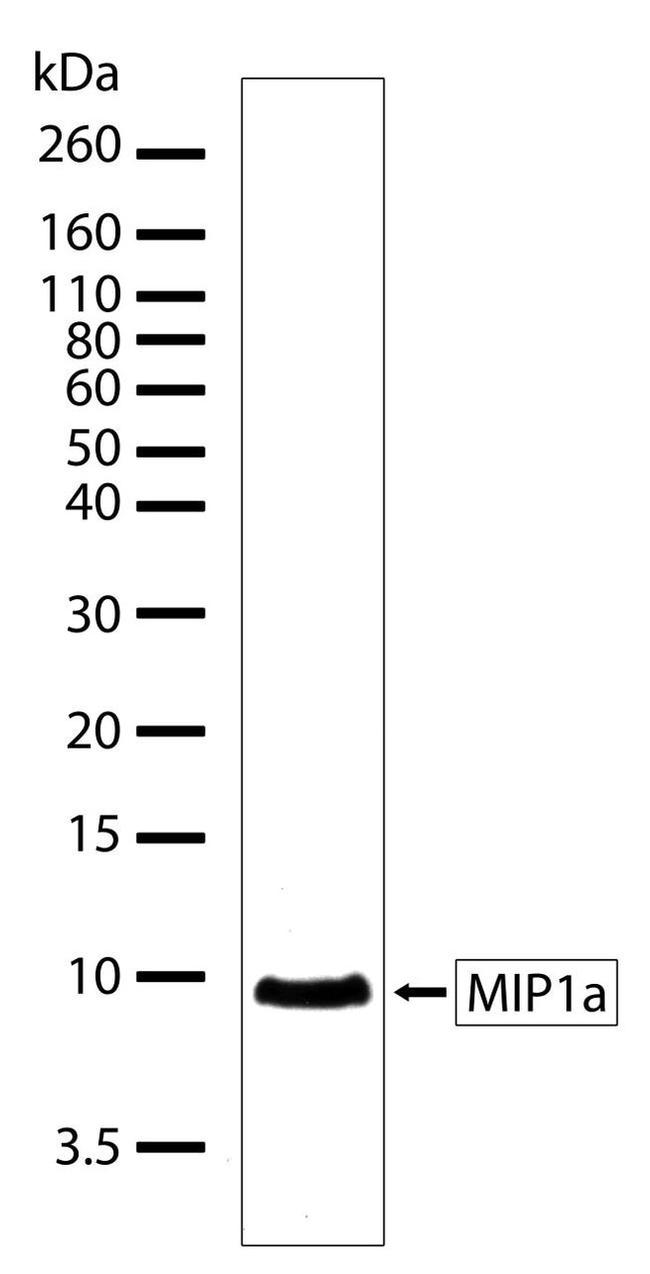
Western blot analysis of active recombinant human MIP1a protein using a MIP-1 alpha recombinant rabbit monoclonal antibody (Product # 701097) at a dilution of 1 µg/mL.