


 全部商品分类
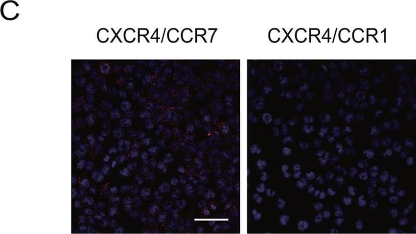


 下载产品说明书 下载SDS
 用小程序,查商品更便捷


 收藏
收藏
 对比
对比 咨询
咨询
Met1-Pro378
Accession # AAA58615
Scientific Data
 View Larger
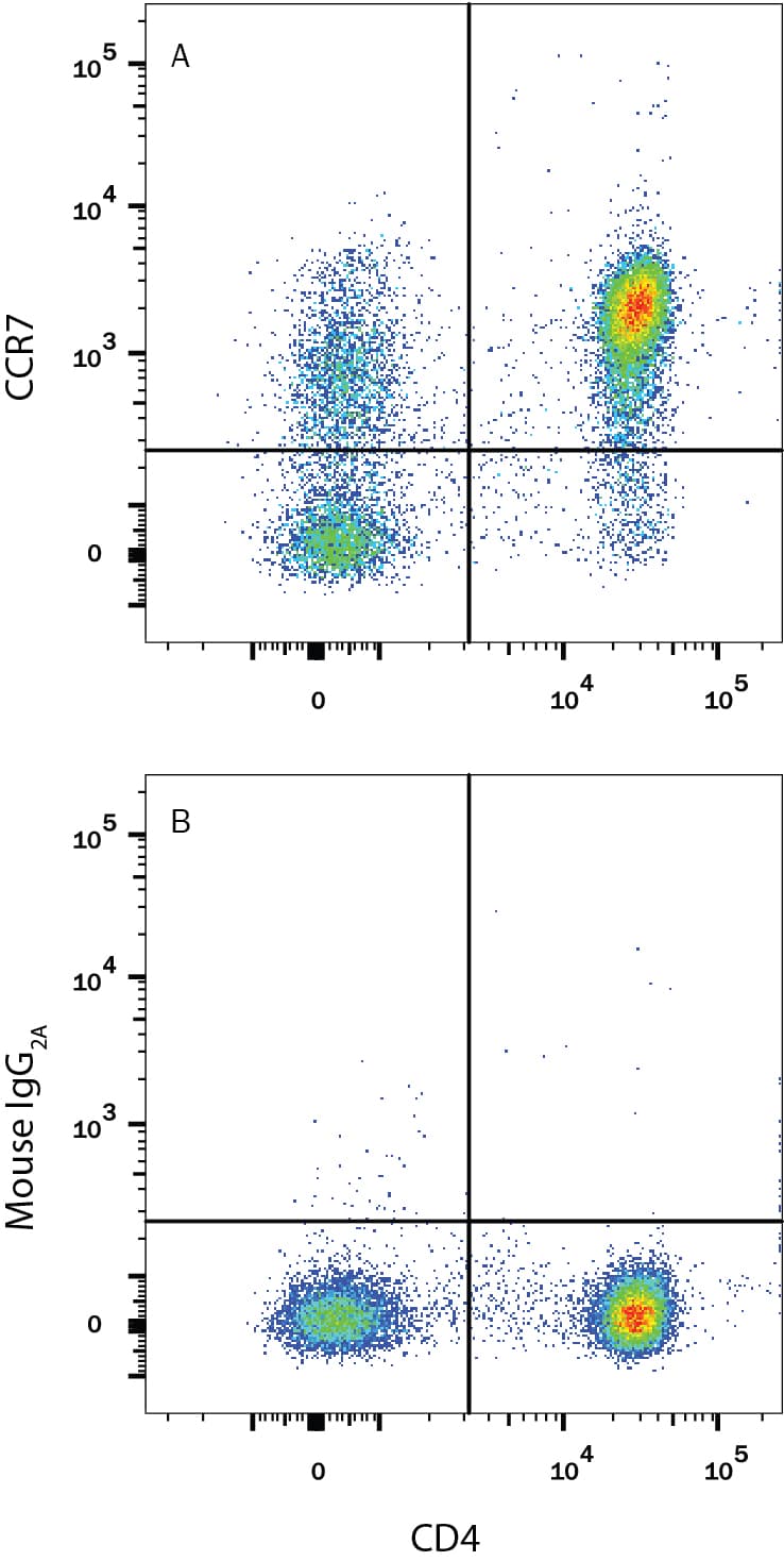
Detection of CCR7 in Human PBMCs by Flow Cytometry. Human peripheral blood mononuclear cells (PBMCs) were stained with Mouse Anti-Human CD4 PE-conjugated Monoclonal Antibody (Catalog # FAB3791P) and either (A) Mouse Anti-Human CCR7 Monoclonal Antibody (Catalog # MAB197) or (B) Mouse IgG2AIsotype Control (Catalog # MAB003) followed by Allophycocyanin-conjugated Anti-Mouse IgG Secondary Antibody (Catalog # F0101B). View our protocol for Staining Membrane-associated Proteins.
.") View Larger
View LargerCCR7 in Human PBMCs. CCR7 was detected in immersion fixed human peripheral blood mononuclear cells (PBMCs) using 25 µg/mL Mouse Anti-Human CCR7 Monoclonal Antibody (Catalog # MAB197) for 3 hours at room temperature. Cells were stained (red) and counterstained (green). View our protocol for Fluorescent ICC Staining of Non-adherent Cells.
.") View Larger
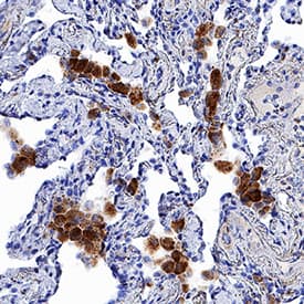
View LargerCCR7 in Human Lung. CCR7 was detected in immersion fixed paraffin-embedded sections of human lung using Mouse Anti-Human CCR7 Monoclonal Antibody (Catalog # MAB197) at 5 µg/mL for 1 hour at room temperature followed by incubation with the Anti-Mouse IgG VisUCyte™ HRP Polymer Antibody (Catalog # VC001). Tissue was stained using DAB (brown) and counterstained with hematoxylin (blue). Specific staining was localized to cytoplasm in macrophages. View our protocol for IHC Staining with VisUCyte HRP Polymer Detection Reagents.
 View Larger
Chemotaxis Induced by CCL19/MIP‑3 beta and Neutral-ization by Human CCR7 Antibody. Recombinant Human CCL19/MIP-3 beta (Catalog # 361-MI) chemoattracts the BaF3 mouse pro-B cell line transfected with human CCR7 in a dose-dependent manner (orange line). The amount of cells that migrated through to the lower chemotaxis chamber was measured by Resazurin (Catalog # AR002). Chemotaxis elicited by Recombinant Human CCL19/MIP-3 beta (50 ng/mL) is neutralized (green line) by increasing concentrations of Mouse Anti-Human CCR7 Monoclonal Antibody (Catalog # MAB197). The ND50 is typically 1-5 µg/mL.
 View Larger
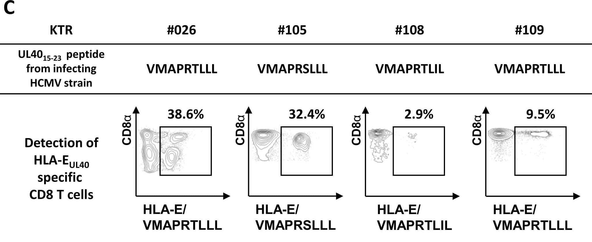
Detection of Human CCR7 by Flow Cytometry HCMV strain-dependent variability of UL4015-23 sequences and HCMV strain-specific HLA-EUL40 T-cell response in hosts.(A, B) Genomic DNAs isolated from HCMV positive blood samples in HCMV+ transplant recipients (n = 25) were sequenced for the identification of UL40 protein (amino acids 1–221) provided by the circulating HCMV strains. (A) Amino acid variability, expressed as a number of amino acid variants, within the HLA-E-binding peptide (UL4015-23, shown in red) among the sequence for HCMV UL40 signal peptide (UL401-37, shown in grey). A total of 32 UL40 sequences from 25 hosts were analysed. UL40 protein sequence from the Merlin HCMV clinical strain was used as reference. Positions 1 to 9 of residues in the HLA-E-binding peptide (UL4015-23) are indicated. (B) Sequence LOGO of the UL4015-23 HLA-E-binding peptide from 25 transplanted hosts. The height of the letter is proportional to the frequency of each amino acid in a given position (P1 to P9). Major anchor residues for binding in the HLA-E peptide groove are indicated in blue. Red letters highlight the important variability observed in position 8 of the HLA-E-binding peptide. Grey boxes correspond to a constitutive deletion of the corresponding amino acid in the UL40 sequence from the infecting viral strain. (C) Representative dot plot analyses showing the detection of strain-specific anti-UL40 HLA-E-restricted CD8 T-cell responses in 4 KTRs (KTR#026, #105, #108 and #109). Frequencies (%) of the HLA-EUL40-specific T cells among total circulating alpha beta CD8 T cells are indicated. Image collected and cropped by CiteAb from the following publication (https://dx.plos.org/10.1371/journal.ppat.1007041), licensed under a CC-BY license. Not internally tested by R&D Systems.
 View Larger
Detection of Human CCR7 by Proximity Ligation Assay CXCR4 ligand binding facilitates CXCR4/CCR7 hetero-oligomer formation.(A) CCR7 homo-oligomer formation in H9 cells after treatment with or without 1 μg/ml recombinant gp120 was examined by in situ PLA. The number of in situ PLA signals per cell was counted by using the Duolink Image Tool software. The result shown is a representative result from three independent experiments showing the mean number of the signals plotted on the vertical axis. *, p < 0.05 by Mann-Whitney’s U test. (B) CCR7 homo-oligomer formation in H9 cells after treatment with 100 ng/ml CXCL12 was examined, as described in (A). Quantification of CCR7 homo-oligomers after CXCL12 pretreatment in the presence or absence of 1 or 5 μg/ml AMD3100. The result shown is a representative result from five independent experiments. *, p < 0.05 by Mann-Whitney’s U test. (C) CXCR4/CCR7 or CXCR4/CCR1 hetero-oligomer formation in H9 cells by in situ PLA using the indicated combinations of antibodies. The z-stack images derived from sections covering 10 μm with a 0.5-μm step are shown. Scale bar represents 50 μm. (D) Quantification of the detected in situ PLA signals per cell was performed. The result shown is a representative result from three independent experiments. *, p < 0.05 by Mann-Whitney’s U test. (E) CXCR4/CCR7 hetero-oligomer formation in H9 cells after treatment with 1 μg/ml gp120 in a native form (N) or control heat-denatured form (D) was examined by the PLA using the indicated combinations of antibodies. (F) CXCR4/CCR7 hetero-oligomer formation was examined with 100 ng/ml CXCL12 as described in (E). Image collected and cropped by CiteAb from the following publication (https://dx.plos.org/10.1371/journal.pone.0117454), licensed under a CC-BY license. Not internally tested by R&D Systems.
 View Larger
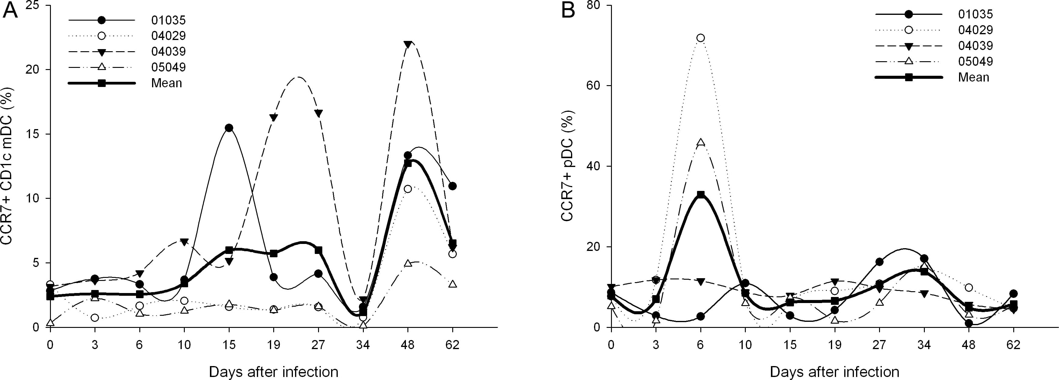
Detection of Rhesus Macaque CCR7 by Flow Cytometry Dynamics of CCR7 expression on CD1c+ mDC and pDC during primary SIVmac239 infection.(A) The dynamics of CCR7 expression on CD1c+ mDC; (B) The dynamics of CCR7 expression on pDC. Image collected and cropped by CiteAb from the following publication (https://dx.plos.org/10.1371/journal.pone.0029036), licensed under a CC-BY license. Not internally tested by R&D Systems.
 View Larger
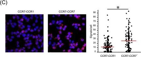
Detection of Human CCR7 by Immunohistochemistry CCR7 homodimers are dissociated by the CCR7 TM4-derived peptide. (A) The levels of bioluminescence signals are presented for HEK293T cells transfected with the indicated combinations of CCR7-CGLuc, CCR7-NGLuc, CGLuc and NGLuc plasmids (1.5 μg each). A representative experiment from at least three independent experiments is shown. Data represent mean ± SD of triplicate samples. *p < 0.05 by one-way ANOVA. (B) The levels of bioluminescence signals are shown for cells transfected with combinations of CCR7-CGLuc and CCR7-NGLuc in the presence of the CCR7 TM4 peptide or the shuffled peptide (15 μg/ml). Relative luminescence was obtained by normalizing the values against untransfected cells. A representative experiment from at least three independent experiments is shown. Data represent mean ± SD of triplicate samples. *p < 0.05 by Student’s t test. (C) The levels of CCR7 homodimer in parental H9 cells were measured by in situ PLA. CCR7-CCR1 heterodimer formation detected by anti-CCR7 mAb-PLAPlus and anti-CCR1 mAb -PLAMinus (left), and CCR7 homodimer formation by anti-CCR7 mAb-PLAPlus and -PLAMinus (right). A representative experiment from at least three independent experiments is shown with the mean number of PLA signals plotted on the vertical axis. *p < 0.05 by Mann-Whitney’s U test. (D) CCR7 (top) or CCR1 (bottom) homodimer formation after treatment with the CCR7 TM4 peptide (right panel) or the shuffled peptide (left panel). The number of PLA signals per cell was counted using the Duolink Image Tool software. A representative experiment from three independent experiments is shown with the mean number of PLA signals plotted on the vertical axis. *p < 0.05 by Mann-Whitney’s U test; NS, not significant. (E) CCR7 expression levels were evaluated in the presence (red open histogram) or absence (blue open histogram) of the CCR7 TM4 peptide (15 μg/ml) by flow cytometry using anti-human CCR7 antibody or isotype control antibody (gray-filled histogram). MFI is indicated on the histograms. A representative experiment from three independent experiments is shown. Image collected and cropped by CiteAb from the following publication (https://www.nature.com/articles/s41598-017-09113-4), licensed under a CC-BY license. Not internally tested by R&D Systems.
 View Larger
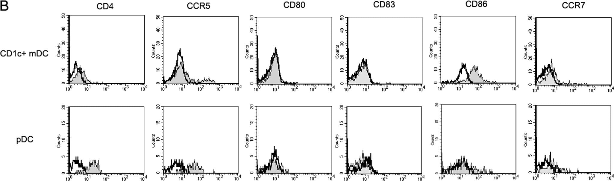
Detection of Rhesus Macaque CCR7 by Flow Cytometry Two-color strategy for identifying CD1c+ mDCs and pDCs.(A) PBMCs (R1) were first gated on Forward-scatter/side-scatter (FSC/SSC) scattergram. Then, CD1c+ mDCs (CD1c+CD14−CD20−) were selected in the R2 region and pDCs (CD123brightHLA-DR+) were selected in the R3 region. (B) The expressions of immunophenotypes, including CD4, CCR5, CD80, CD83, CD86, and CCR7 on DCs subsets were analyzed. Open histograms correspond to isotype controls and filled gray histograms correspond to specific mAbs staining. Image collected and cropped by CiteAb from the following publication (https://dx.plos.org/10.1371/journal.pone.0029036), licensed under a CC-BY license. Not internally tested by R&D Systems.
 View Larger
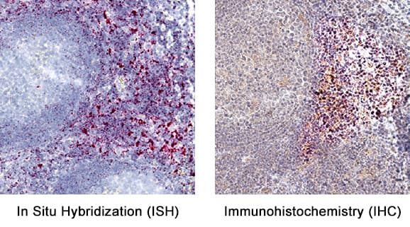
Detection of CCR7 in Human Tonsil. Formalin-fixed paraffin-embedded tissue sections of human tonsil were probed for CCR7 mRNA (ACD RNAScope Probe, catalog # 410728; Fast Red chromogen, ACD catalog # 322750). Adjacent tissue section was processed for immunohistochemistry using mouse anti-human CCR7 monoclonal antibody (R&D Systems catalog # MAB197) at 15ug/mL with overnight incubation at 4 degrees Celsius followed by incubation with anti-mouse IgG VisUCyte HRP Polymer Antibody (Catalog # VC001) and DAB chromogen (yellow-brown). Tissue was counterstained with hematoxylin (blue). Specific staining was localized to lymphocytes.
Human CCR7 Antibody Summary
Met1-Pro378
Accession # AAA58615
*Small pack size (-SP) is supplied either lyophilized or as a 0.2 µm filtered solution in PBS.
Applications
Please Note: Optimal dilutions should be determined by each laboratory for each application. General Protocols are available in the Technical Information section on our website.
Background: CCR7
CCR7 (Chemokine Receptor 7; also CD197) is a 7 transmembrane (7TM) G protein-coupled chemokine receptor for the homeostatic chemokines CCL19/MIP-3 beta and CCL21/6Ckine. CCL19 and CCL21 are constitutively expressed by high endothelial venule epithelial cells or fibroblastic reticular cells in secondary lymphoid organs. CCR7 is upregulated on dendritic cells, naïve and memory T cells, Treg, NK cells, and B cells following inflammatory stimulation. Its expression enables the function of immune cell trafficking to and retention in regional lymph nodes for expansion of the adaptive immune response. Human CCR7 shares 87% amino acid sequence identity with mouse CCR7.
Preparation and Storage
- 12 months from date of receipt, -20 to -70 °C as supplied.
- 1 month, 2 to 8 °C under sterile conditions after reconstitution.
- 6 months, -20 to -70 °C under sterile conditions after reconstitution.
参考图片
CCR7 in Human PBMCs.
CCR7 was detected in immersion fixed human peripheral blood mononuclear cells (PBMCs) using 25 µg/mL Mouse Anti-Human CCR7 Monoclonal Antibody (Catalog # MAB197) for 3 hours at room temperature. Cells were stained (red) and counterstained (green). View our protocol for Fluorescent ICC Staining of Non-adherent Cells.
Detection of CCR7 in Human PBMCs by Flow Cytometry. Human peripheral blood mononuclear cells (PBMCs) were stained with Mouse Anti-Human CD4 PE‑conjugated Monoclonal Antibody (Catalog # FAB3791P) and either (A) Mouse Anti-Human CCR7 Monoclonal Antibody (Catalog # MAB197) or (B) Mouse IgG2A Isotype Control (Catalog # MAB003) followed by Allophycocyanin-conjugated Anti-Mouse IgG Secondary Antibody (Catalog # F0101B). View our protocol for Staining Membrane-associated Proteins.
Chemotaxis Induced by CCL19/MIP‑3 beta and Neutralization by Human CCR7 Antibody.
Recombinant Human CCL19/MIP‑3 beta (Catalog # 361‑MI) chemoattracts the BaF3 mouse pro‑B cell line transfected with human CCR7 in a dose-dependent manner (orange line). The amount of cells that migrated through to the lower chemotaxis chamber was measured by Resazurin (Catalog # AR002). Chemotaxis elicited by Recombinant Human CCL19/MIP‑3 beta (50 ng/mL) is neutralized (green line) by increasing concentrations of Mouse Anti-Human CCR7 Monoclonal Antibody (Catalog # MAB197). The ND50 is typically 1-5 µg/mL.