


 全部商品分类
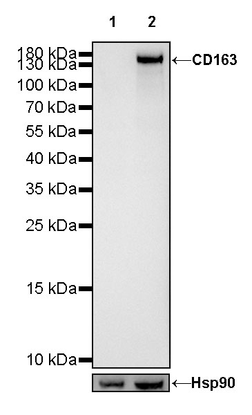


S-RMab® CD163 Recombinant Rabbit mAb (SDT-R164)

 下载产品说明书
 用小程序,查商品更便捷


 收藏
收藏
 对比
对比 咨询
咨询
WB
1:500IHC-P
1:1000-1:2000IF
1:2000IP
1:25
CD163 is a monocyte/macrophage specific marker expressed predominantly on cells which possess strong anti-inflammatory potential. The expression of CD163 is strongly induced by anti-inflammatory mediators such as glucocorticoids and interleukin-10, while being inhibited by pro-inflammatory mediators such as interferon-gamma. CD163-expressing mononuclear phagocytes, as well as soluble CD163, may both take part in downregulating an inflammatory response [PubMed: 22038213].
参考图片
WB result of CD163 Rabbit mAb
Primary antibody: CD163 Rabbit mAb at 1/500 dilution
Lane 1: rat heart lysate 20 µg
Lane 2: rat spleen lysate 20 µg
Negative control: rat heart lysate
Secondary antibody: Goat Anti-Rabbit IgG, (H+L), HRP conjugated at 1/10000 dilution
Predicted MW: 125kDa
Observed MW: 160kDa
Exposure time: 180s
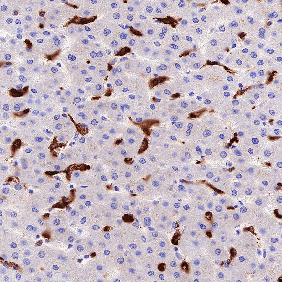
IHC shows positive staining in paraffin-embedded human liver. Anti-CD163 antibody was used at 1/2000 dilution, followed by a HRP Polymer for Mouse & Rabbit IgG (ready to use). Counterstained with hematoxylin. Heat mediated antigen retrieval with Tris/EDTA buffer pH9.0 was performed before commencing with IHC staining protocol.
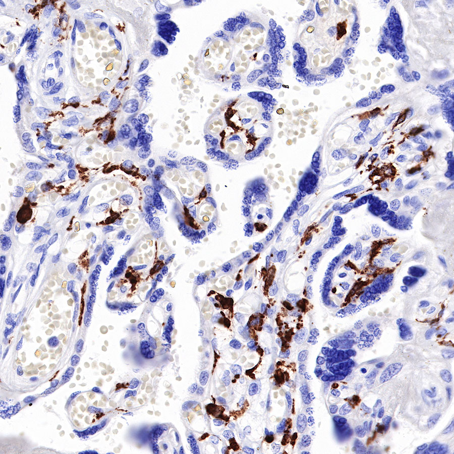
IHC shows positive staining in paraffin-embedded human placenta. Anti-CD163 antibody was used at 1/1000 dilution, followed by a HRP Polymer for Mouse & Rabbit IgG (ready to use). Counterstained with hematoxylin. Heat mediated antigen retrieval with Tris/EDTA buffer pH9.0 was performed before commencing with IHC staining protocol.
IHC shows positive staining in paraffin-embedded human spleen. Anti-CD163 antibody was used at 1/1000 dilution, followed by a HRP Polymer for Mouse & Rabbit IgG (ready to use). Counterstained with hematoxylin. Heat mediated antigen retrieval with Tris/EDTA buffer pH9.0 was performed before commencing with IHC staining protocol.
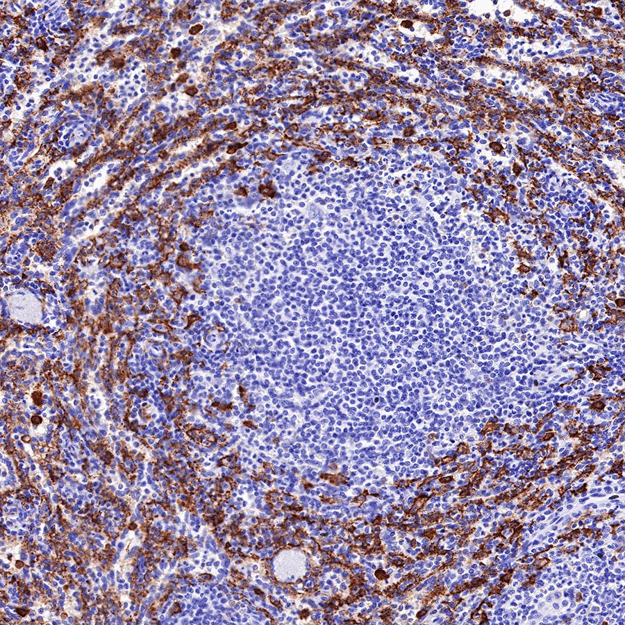
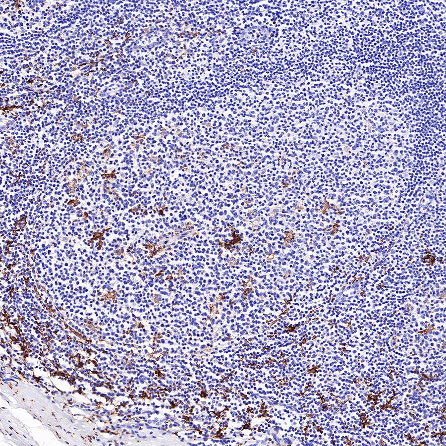
IHC shows positive staining in paraffin-embedded human tonsil. Anti-CD163 antibody was used at 1/1000 dilution, followed by a HRP Polymer for Mouse & Rabbit IgG (ready to use). Counterstained with hematoxylin. Heat mediated antigen retrieval with Tris/EDTA buffer pH9.0 was performed before commencing with IHC staining protocol.
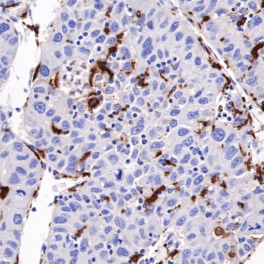
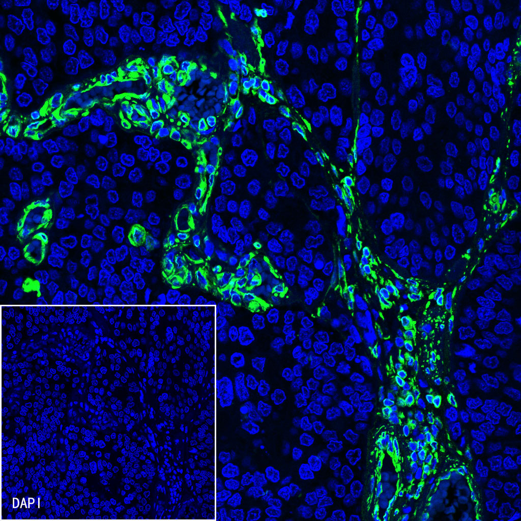
IHC shows positive staining in paraffin-embedded human hepatocellular carcinoma. Anti-CD163 antibody was used at 1/1000 dilution, followed by a HRP Polymer for Mouse & Rabbit IgG (ready to use). Counterstained with hematoxylin. Heat mediated antigen retrieval with Tris/EDTA buffer pH9.0 was performed before commencing with IHC staining protocol.
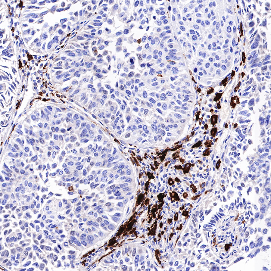
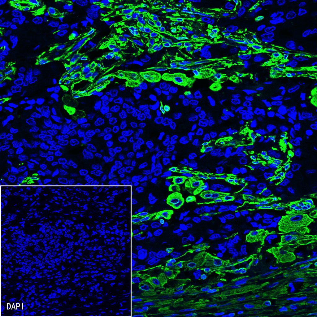
IHC shows positive staining in paraffin-embedded human lung squamous cell carcinoma. Anti-CD163 antibody was used at 1/1000 dilution, followed by a HRP Polymer for Mouse & Rabbit IgG (ready to use). Counterstained with hematoxylin. Heat mediated antigen retrieval with Tris/EDTA buffer pH9.0 was performed before commencing with IHC staining protocol.
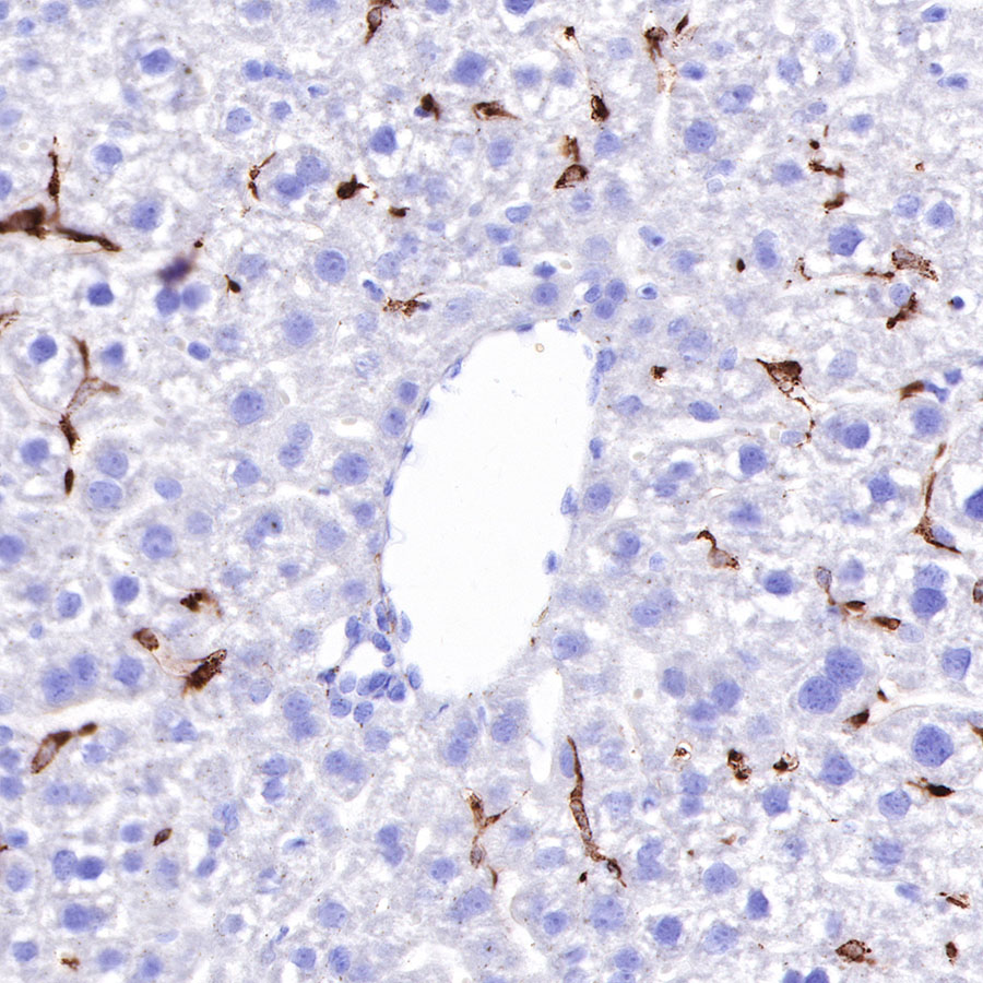
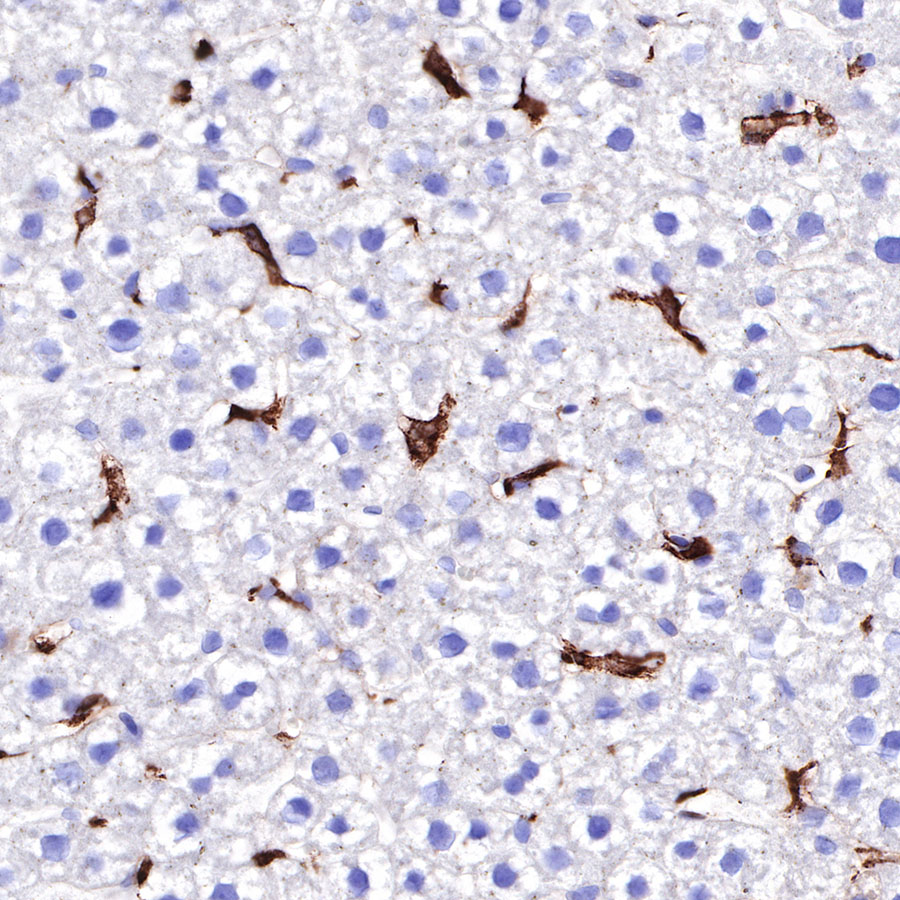
IHC shows positive staining in paraffin-embedded mouse liver. Anti-CD163 antibody was used at 1/1000 dilution, followed by a HRP Polymer for Mouse & Rabbit IgG (ready to use). Counterstained with hematoxylin. Heat mediated antigen retrieval with Tris/EDTA buffer pH9.0 was performed before commencing with IHC staining protocol.
IHC shows positive staining in paraffin-embedded rat liver. Anti-CD163 antibody was used at 1/1000 dilution, followed by a HRP Polymer for Mouse & Rabbit IgG (ready to use). Counterstained with hematoxylin. Heat mediated antigen retrieval with Tris/EDTA buffer pH9.0 was performed before commencing with IHC staining protocol.
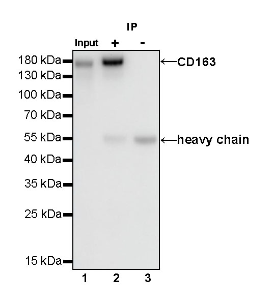
CD163 Rabbit mAb at 1/25 dilution (1 µg) immunoprecipitating CD163 in 0.4 mg rat spleen whole cell lysate.
Western blot was performed on the immunoprecipitate using CD163 Rabbit mAb at 1/1000 dilution.
Secondary antibody (HRP) for IP was used at 1/400 dilution.
Lane 1: rat spleen whole cell lysate 20 µg (Input)
Lane 2: CD163 Rabbit mAb IP in rat spleen whole cell lysate
Lane 3: Rabbit monoclonal IgG IP in rat spleen whole cell lysate
Predicted MW: 125 kDa
Observed MW: 160 kDa
Exposure time: 180 s