The F10/21A3 monoclonal antibody specifically binds to CD1c. The CD1 family of transmembrane glycoproteins are structurally related to the classical major histocompatibility complex (MHC) proteins. CD1c is a type I transmembrane glycoprotein that forms heterodimers with beta-2-microglobulin. CD1c presents lipids and glycolipids of self or microbial origin to T cells. CD1c is expressed by Langerhans cells, dendritic cells, monocytes, cortical thymocytes, T cells, and some B cells.
The antibody was conjugated to BD Horizon BV421 which is part of the BD Horizon Brilliant™ Violet family of dyes. With an Ex Max of 407-nm and Em Max at 421-nm, BD Horizon BV421 can be excited by the violet laser and detected in the standard Pacific Blue™ filter set (eg, 450/50-nm filter). BD Horizon BV421 conjugates are very bright, often exhibiting a 10 fold improvement in brightness compared to Pacific Blue conjugates.
商品描述
F10/21A3
The F10/21A3 monoclonal antibody specifically binds to CD1c. The CD1 family of transmembrane glycoproteins are structurally related to the classical major histocompatibility complex (MHC) proteins. CD1c is a type I transmembrane glycoprotein that forms heterodimers with beta-2-microglobulin. CD1c presents lipids and glycolipids of self or microbial origin to T cells. CD1c is expressed by Langerhans cells, dendritic cells, monocytes, cortical thymocytes, T cells, and some B cells.
The antibody was conjugated to BD Horizon BV421 which is part of the BD Horizon Brilliant™ Violet family of dyes. With an Ex Max of 407-nm and Em Max at 421-nm, BD Horizon BV421 can be excited by the violet laser and detected in the standard Pacific Blue™ filter set (eg, 450/50-nm filter). BD Horizon BV421 conjugates are very bright, often exhibiting a 10 fold improvement in brightness compared to Pacific Blue conjugates.
同种型
Mouse IgG1, κ
克隆号
克隆 F10/21A3 (RUO)
产品详情
BV421
The BD Horizon Brilliant Violet™ 421 (BV421) Dye is part of the BD Horizon Brilliant Violet™ family of dyes. This polymer-technology based dye has an excitation maximum (Ex Max) of 407-nm and an emission maximum (Em Max) at 423-nm. Driven by BD innovation, BV421 is designed to be excited by the violet laser (405-nm) and detected using an optical filter centered near 420-nm (e.g., a 431/28-nm or 450/50-nm bandpass filter). BV421 is an ideal alternative for V450 as it is approximately ten times brighter with less spillover into the BV510/V500 detector. Please ensure that your instrument’s configurations (lasers and optical filters) are appropriate for this dye.
BV421
Violet 405 nm
407 nm
423 nm
应用
实验应用
Flow cytometry (Routinely Tested)
推荐用量
5 µl
反应种属
Human (QC Testing)
目标/特异性
CD1c
背景
别名
CD1; R7; M241; BDCA1
制备和贮存
存储溶液
Aqueous buffered solution containing BSA and ≤0.09% sodium azide.
保存方式
Aqueous buffered solution containing BSA and ≤0.09% sodium azide.
文献
文献
研发参考(3)
1. Delia D, Cattoretti G, Polli N, et al. CD1c but neither CD1a nor CD1b molecules are expressed on normal, activated, and malignant human B cells: identification of a new B-cell subset. Blood. 1988; 72(1):241-247. (Biology).
2. Grant EP, Degano M, Rosat JP, et al. Molecular recognition of lipid antigens by T cell receptors. J Exp Med. 1999; 189(1):195-205. (Clone-specific: Blocking, Functional assay, Inhibition).
3. Moody DB, Ulrichs T, Mühlecker W, et al. CD1c-mediated T-cell recognition of isoprenoid glycolipids in Mycobacterium tuberculosis infection. Nature. 2000; 404(6780):884-888. (Clone-specific: Blocking, Functional assay, Inhibition).
数据库链接
Entrez-Gene ID
911
参考图片
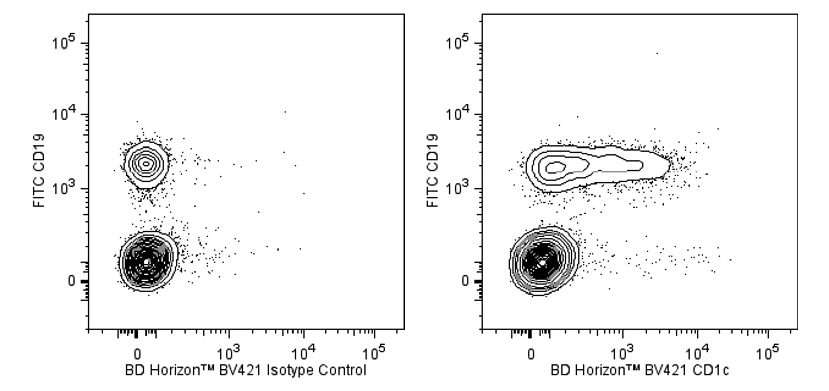
Two-color flow cytometric analysis of CD1c expression on human peripheral blood lymphocytes. Human whole blood was stained with FITC Mouse Anti-Human CD19 antibody (Cat. No. 555412/560994) and either BD Horizon™ BV421 Mouse IgG1, κ Isotype Control (Cat. No. 562438; Left Panel) or BD Horizon™ BV421 Mouse Anti-Human CD1c (Cat. No. 565050/565051; Right Panel). Erythrocytes were lysed with BD FACS Lysing Solution (Cat. No. 349202). Two-color flow cytometric contour plots showing the correlated expression of CD1c (or Ig Isotype control staining) versus CD19 were derived from gated events with the forward and side light-scatter characteristics of intact lymphocytes. Flow cytometric analysis was performed using a BD™ LSR II Flow Cytometer System.
Two-color flow cytometric analysis of CD1c expression on human peripheral blood lymphocytes. Human whole blood was stained with FITC Mouse Anti-Human CD19 antibody (Cat. No. 555412/560994) and either BD Horizon™ BV421 Mouse IgG1, κ Isotype Control (Cat. No. 562438; Left Panel) or BD Horizon™ BV421 Mouse Anti-Human CD1c (Cat. No. 565050/565051; Right Panel). Erythrocytes were lysed with BD FACS Lysing Solution (Cat. No. 349202). Two-color flow cytometric contour plots showing the correlated expression of CD1c (or Ig Isotype control staining) versus CD19 were derived from gated events with the forward and side light-scatter characteristics of intact lymphocytes. Flow cytometric analysis was performed using a BD™ LSR II Flow Cytometer System.


 全部商品分类


 下载产品说明书
 用小程序,查商品更便捷


 收藏
收藏
 对比
对比 咨询
咨询
