The F10/21A3 monoclonal antibody specifically binds to CD1c. The CD1 family of transmembrane glycoproteins are structurally related to the classical major histocompatibility complex (MHC) proteins. CD1c is a type I transmembrane glycoprotein that forms heterodimers with beta-2-microglobulin. CD1c presents lipids and glycolipids of self or microbial origin to T cells. CD1c is expressed by Langerhans cells, dendritic cells, monocytes, cortical thymocytes, T cells, and some B cells.
This antibody was conjugated to BD Horizon APC-R700, which has been developed exclusively by BD Biosciences as a better alternative to Alexa Fluor® 700. APC-R700 excites and emits at similar wavelengths to Alexa Fluor® 700 yet exhibits significantly improved brightness. This dye can be excited by the red laser and detected with the same filter set as Alexa Fluor® (eg, 730/45-nm filter).
商品描述
F10/21A3
The F10/21A3 monoclonal antibody specifically binds to CD1c. The CD1 family of transmembrane glycoproteins are structurally related to the classical major histocompatibility complex (MHC) proteins. CD1c is a type I transmembrane glycoprotein that forms heterodimers with beta-2-microglobulin. CD1c presents lipids and glycolipids of self or microbial origin to T cells. CD1c is expressed by Langerhans cells, dendritic cells, monocytes, cortical thymocytes, T cells, and some B cells.
This antibody was conjugated to BD Horizon APC-R700, which has been developed exclusively by BD Biosciences as a better alternative to Alexa Fluor® 700. APC-R700 excites and emits at similar wavelengths to Alexa Fluor® 700 yet exhibits significantly improved brightness. This dye can be excited by the red laser and detected with the same filter set as Alexa Fluor® (eg, 730/45-nm filter).
同种型
Mouse IgG1, κ
克隆号
克隆 F10/21A3 (RUO)
产品详情
APC-R700
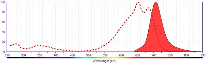
The BD Horizon™ APC-R700 (APC-R700) Dye is a part of the BD APC red family of dyes. This tandem fluorochrome is comprised of an Allophycocyanin (APC) dye donor that has excitation maximum (Ex Max) of 651-nm and an acceptor dye, R700, with an emission maximum (Em Max) at 706-nm. APC-R700, driven by BD innovation, is designed to be excited by the red (627–640-nm) laser and detected using an optical filter centered near 710-nm (e.g., a 720/40-nm bandpass filter). APC-R700 is a brighter alternative to Alexa Fluor™ 700. Please ensure that your instrument’s configurations (lasers and optical filters) are appropriate for this dye.
APC-R700
Red 627-640 nm
651 nm
706 nm
应用
实验应用
Flow cytometry (Routinely Tested)
推荐用量
5 µl
反应种属
Human (QC Testing)
目标/特异性
CD1c
背景
别名
CD1; R7; M241; BDCA1
制备和贮存
存储溶液
Aqueous buffered solution containing BSA, protein stabilizer, glycerol and ≤0.09% sodium azide.
保存方式
Aqueous buffered solution containing BSA, protein stabilizer, glycerol and ≤0.09% sodium azide.
文献
文献
研发参考(4)
1. Delia D, Cattoretti G, Polli N, et al. CD1c but neither CD1a nor CD1b molecules are expressed on normal, activated, and malignant human B cells: identification of a new B-cell subset. Blood. 1988; 72(1):241-247. (Biology).
2. Grant EP, Degano M, Rosat JP, et al. Molecular recognition of lipid antigens by T cell receptors. J Exp Med. 1999; 189(1):195-205. (Clone-specific: Blocking, Functional assay, Inhibition).
3. Melian A, Geng YJ, Sukhova GK, Libby P, Porcelli SA. CD1 expression in human atherosclerosis. A potential mechanism for T cell activation by foam cells. Am J Pathol. 1999; 155(3):775-786. (Immunogen: Flow cytometry).
4. Moody DB, Ulrichs T, Mühlecker W, et al. CD1c-mediated T-cell recognition of isoprenoid glycolipids in Mycobacterium tuberculosis infection. Nature. 2000; 404(6780):884-888. (Clone-specific: Blocking, Functional assay, Inhibition).
数据库链接
Entrez-Gene ID
911
参考图片
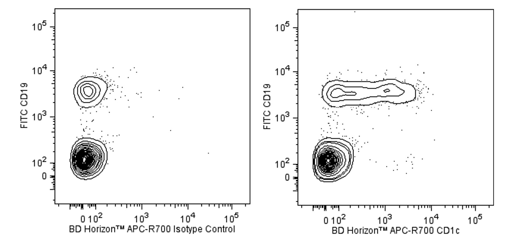
Two-color flow cytometric analysis of CD1c expression on human peripheral blood lymphocytes. Human whole blood was stained with FITC Mouse Anti-Human CD19 antibody (Cat. No. 555412/560994) and either BD Horizon™ APC-R700 Mouse IgG1, κ Isotype Control (Cat. No. 564974; Left Plot) or BD Horizon™ APC-R700 Mouse Anti-Human CD1c antibody (Cat. No. 566614/566615; Right Plot). Erythrocytes were lysed with BD FACS Lysing Solution (Cat. No. 349202). Two-color flow cytometric contour plots showing the correlated expression of CD1c (or Ig Isotype control staining) versus CD19 were derived from gated events with the forward and side light-scatter characteristics of intact lymphocytes. Flow cytometric analysis was performed using a BD LSRFortessa™ Cell Analyzer System.


 全部商品分类


 下载产品说明书
 用小程序,查商品更便捷


 收藏
收藏
 对比
对比 咨询
咨询