


 全部商品分类


PD-L1 (E1L3N®) XP® Rabbit mAb

 下载产品说明书 下载COA 下载SDS
 用小程序,查商品更便捷


 收藏
收藏
 对比
对比 咨询
咨询
Monoclonal antibody is produced by immunizing animals with a synthetic peptide corresponding to residues near the carboxy terminus of human PD-L1 protein.
Product Usage Information
| Application | Dilution |
|---|---|
| Western Blotting | 1:1000 |
| Immunoprecipitation | 1:50 |
| IHC Leica Bond | 1:200 - 1:800 |
| Immunohistochemistry (Paraffin) | 1:100 - 1:400 |
| Flow Cytometry (Fixed/Permeabilized) | 1:200 - 1:800 |
Specificity/Sensitivity
Species Reactivity:
Human
Supplied in 10 mM sodium HEPES (pH 7.5), 150 mM NaCl, 100 µg/ml BSA, 50% glycerol and less than 0.02% sodium azide. Store at –20°C. Do not aliquot the antibody.
For a carrier free (BSA and azide free) version of this product see product #85164.
参考图片
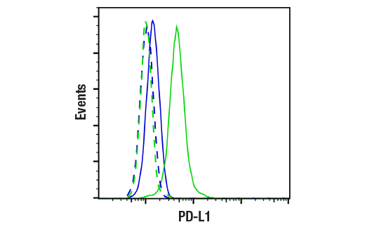
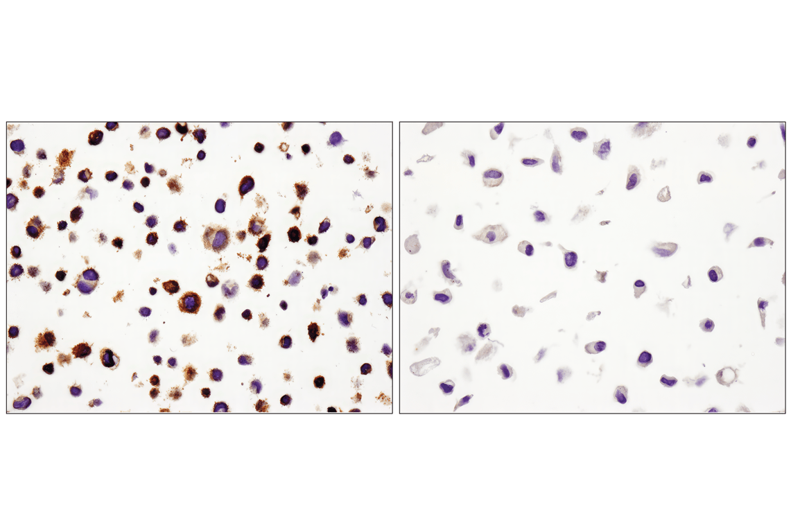
Flow cytometric analysis of Ramos cells (blue, negative) and Sup-M2 cells (green, positive) using PD-L1 (E1L3N®) XP® Rabbit mAb (solid lines) or concentration-matched Rabbit (DA1E) mAb IgG XP® Isotype Control #3900 (dashed lines). Anti-rabbit IgG (H+L), F(ab')2 Fragment (Alexa Fluor® 488 Conjugate) #4412 was used as a secondary antibody.
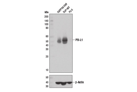
Western blot analysis of extracts from KARPAS-299, SUP-M2, and PC-3 cells using PD-L1 (E1L3N®) XP® Rabbit mAb (upper) and β-Actin (D6A8) Rabbit mAb #8457 (lower).
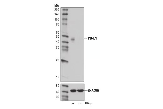
Western blot analysis of extracts from A549 cells, IFN-γ treated (100 ng/mL, 48 hr; +) or untreated (-), using PD-L1 (E1L3N®) XP® Rabbit mAb (upper) or β-Actin (D6A8) Rabbit mAb #8457 (lower).
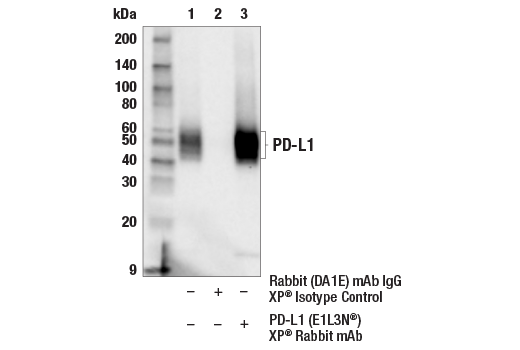
Immunoprecipitation of PD-L1 protein from KARPAS-299 extracts. Lane 1 is 10% input, lane 2 is Rabbit (DA1E) mAb IgG XP® Isotype Control #3900, and lane 3 is PD-L1 (E1L3N®) XP® Rabbit mAb. Western blot analysis was performed using PD-L1 (405.9A11) Mouse mAb #29122. Anti-mouse IgG, HRP-linked Antibody #7076 was used as a secondary antibody.
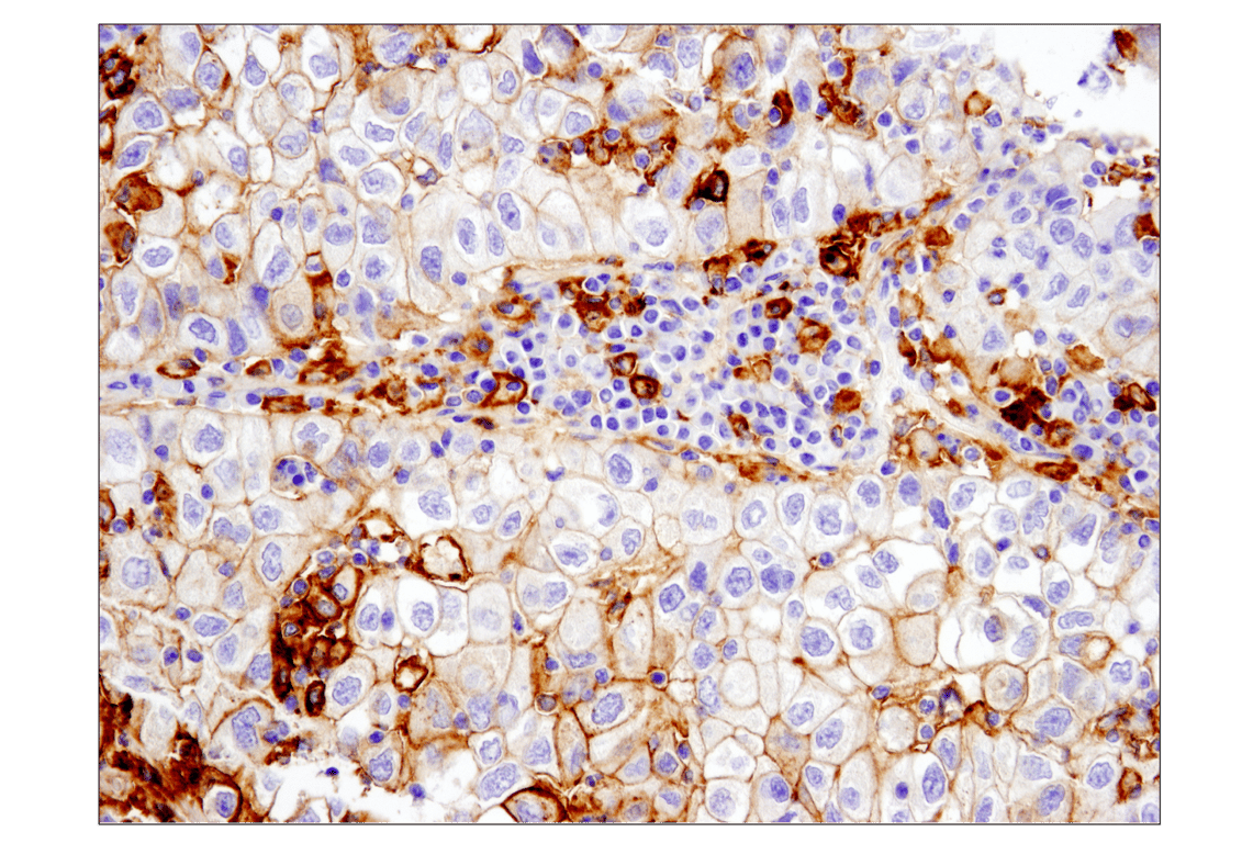
Immunohistochemical analysis of paraffin-embedded human non-small cell lung carcinoma using PD-L1 (E1L3N®) XP® Rabbit mAb performed on the Leica® BOND™ Rx.
Immunohistochemical analysis of paraffin-embedded human lung carcinoma using PD-L1 (E1L3N®) XP® Rabbit mAb.
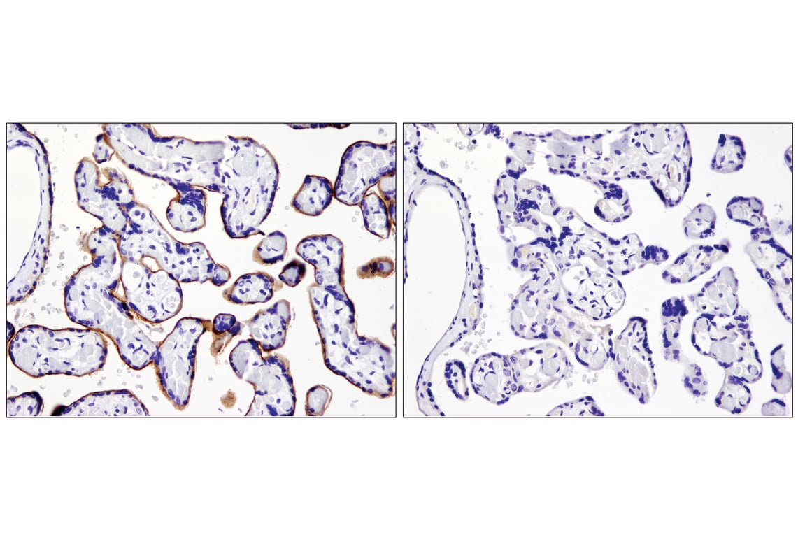
Immunohistochemical analysis of paraffin-embedded human placenta using PD-L1 (E1L3N®) XP® Rabbit mAb in the presence of control peptide (left) or antigen-specific peptide (right).
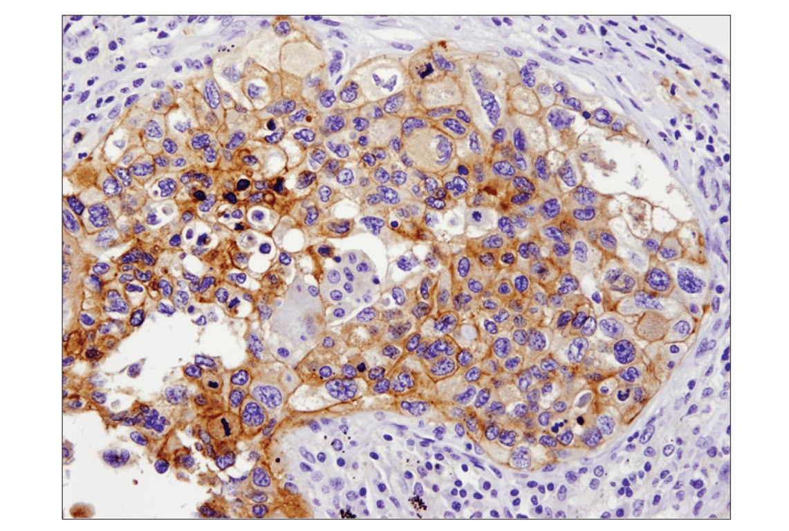
Immunohistochemical analysis of paraffin-embedded Karpas-299 (left) or PC-3 (right) cell pellets using PD-L1 (E1L3N®) XP® Rabbit mAb.
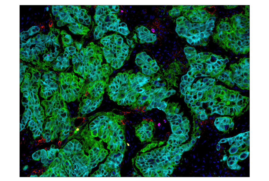
Multiplex immunohistochemical analysis of paraffin-embedded human ovarian carcinoma using PD-L1 (E1L3N®) XP® rabbit mAb (red), CD8α (C8/144B) mouse mAb #70306 (magenta), B7-H4 (D1M8I) XP® rabbit mAb #14572 (green), FoxP3 (D2W8E™) rabbit mAb #98377 (yellow), and Pan-keratin (C11) mouse mAb #4545 (cyan).
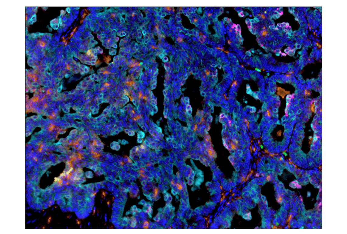
Multiplex immunohistochemical analysis of paraffin-embedded human ovarian carcinoma using PD-L1 (E1L3N®) XP® rabbit mAb (red), PD-L2 (D7U8C™) XP® rabbit mAb (magenta) #82723, Arginase-1 (D4E3M™) XP® rabbit mAb #93668 (green), IDO (D5J4E™) rabbit mAb #86630 (yellow), CD68 (D4B9C) XP® rabbit mAb #76437 (orange), and Pan-keratin (C11) mouse mAb #4545 (cyan).
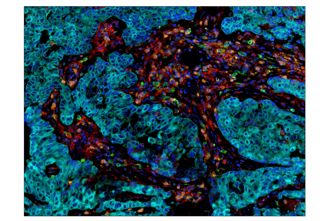
Multiplex immunohistochemical analysis of paraffin-embedded human breast carcinoma using PD-L1 (E1L3N®) XP® rabbit mAb (red), PD-1 (D4W2J) XP® rabbit mAb #86163 (green), LAG3 (D2G4O™) XP® rabbit mAb #15372 (magenta), TIM-3 (D5D5R™) XP® rabbit mAb #45208 (yellow), CD8α (C8/144B) mouse mAb #70306 (orange), and Pan-keratin (C11) mouse mAb #4545 (cyan).