


 全部商品分类


 用小程序,查商品更便捷


Monoclonal antibody is produced by immunizing animals with a synthetic peptide corresponding to residues near the carboxy terminus of human PD-L1 protein.
Product Usage Information
| Application | Dilution |
|---|---|
| Western Blotting | 1:1000 |
| Simple Western™ | 1:10 - 1:50 |
| Immunohistochemistry (Paraffin) | 1:100 - 1:400 |
Specificity/Sensitivity
Species Reactivity:
Human
Supplied in 10 mM sodium HEPES (pH 7.5), 150 mM NaCl, 100 µg/ml BSA, 50% glycerol and less than 0.02% sodium azide. Store at –20°C. Do not aliquot the antibody.
For a carrier-free (BSA and azide free) version of this product see product #39356.
参考图片
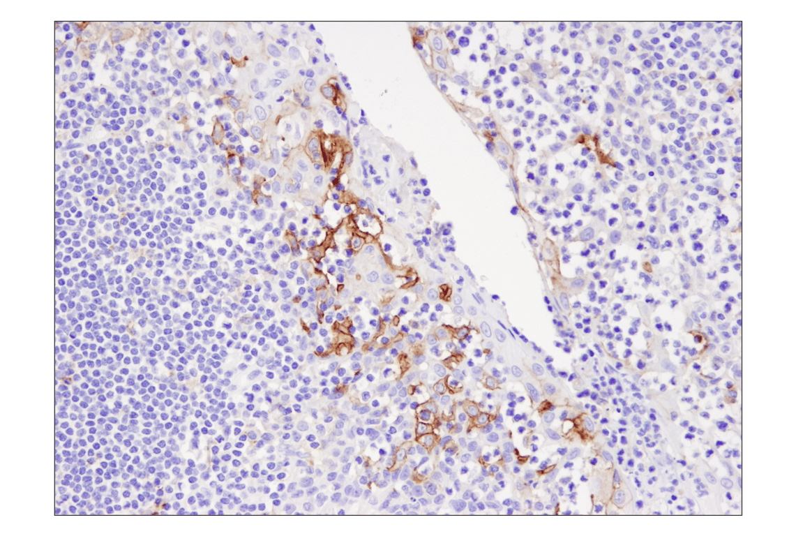
Immunohistochemical analysis of parffin-embedded human tonsil using PD-L1 (405.9A11) Mouse mAb.
Western blot analysis of extracts from various cell lines using PD-L1 (405.9A11) Mouse mAb (upper) or β-Actin (D6A8) Rabbit mAb #8457 (lower). KARPAS-299 cell line source: Dr Abraham Karpas, University of Cambridge.
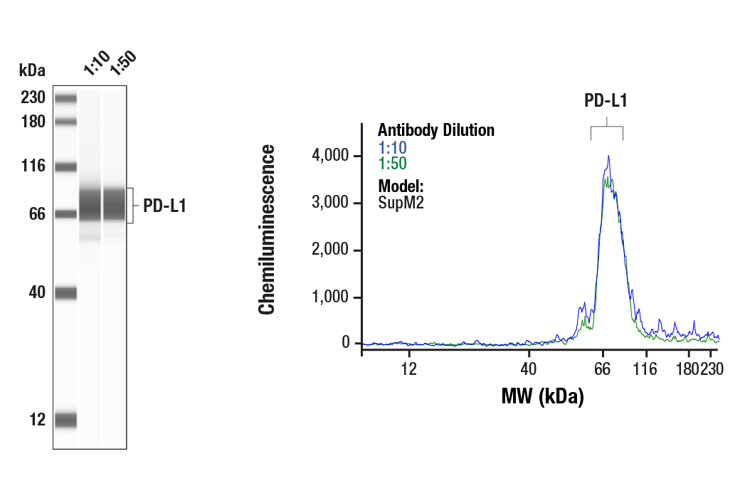
Simple Western™ analysis of SupM2 lysates (0.1 mg/mL) using PD-L1 (405.9A11) Mouse mAb #29122. The virtual lane view (left) shows the target band (as indicated) at 1:10 and 1:50 dilutions of primary antibody. The corresponding electropherogram view (right) plots chemiluminescence by molecular weight along the capillary at 1:10 (blue line) and 1:50 (green line) dilutions of primary antibody. This experiment was performed under reducing conditions on the Jess™ Simple Western instrument from ProteinSimple, a BioTechne brand, using the 12-230 kDa separation module.
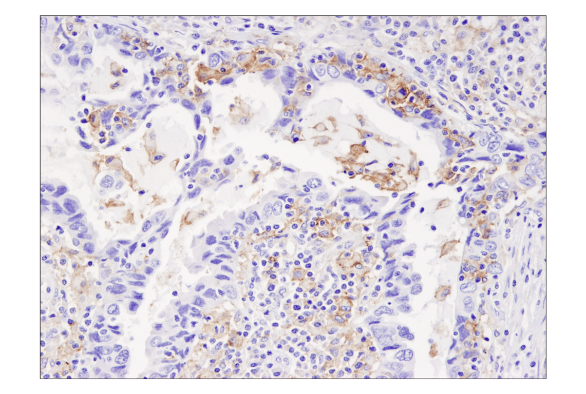
Immunohistochemical analysis of paraffin-embedded human breast carcinoma using PD-L1 (405.9A11) Mouse mAb.
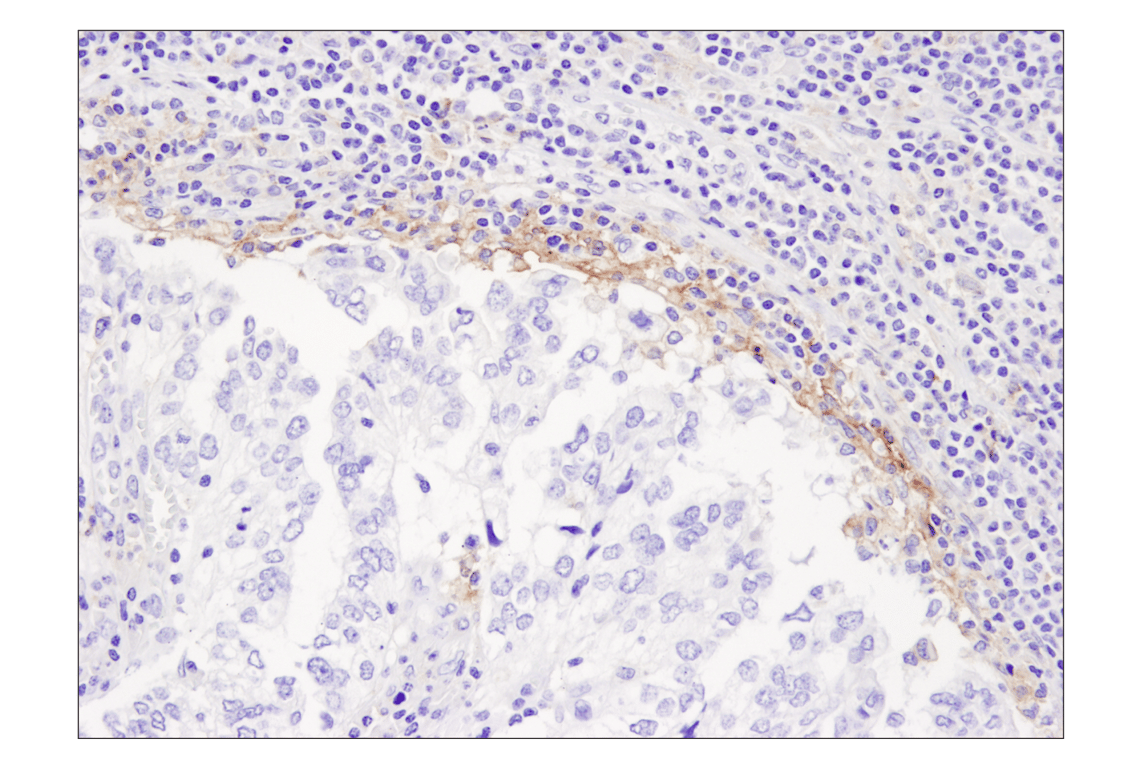
Immunohistochemical analysis of paraffin-embedded human lung carcinoma using PD-L1 (405.9A11) Mouse mAb.
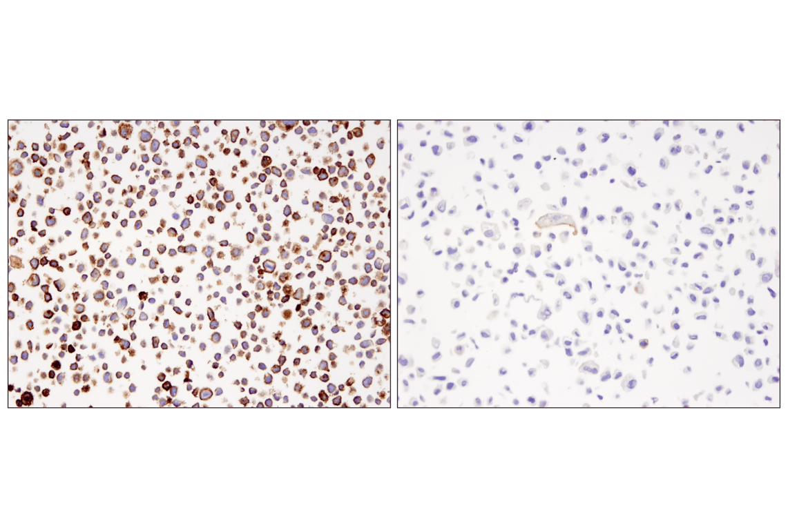
Immunohistochemical analysis of paraffin-embedded HDLM-2 cell pellet (left) or PC-3 cell pellet (right) using PD-L1 (405.9A11) Mouse mAb on SignalSlide® PD-L1 IHC Controls #13747.
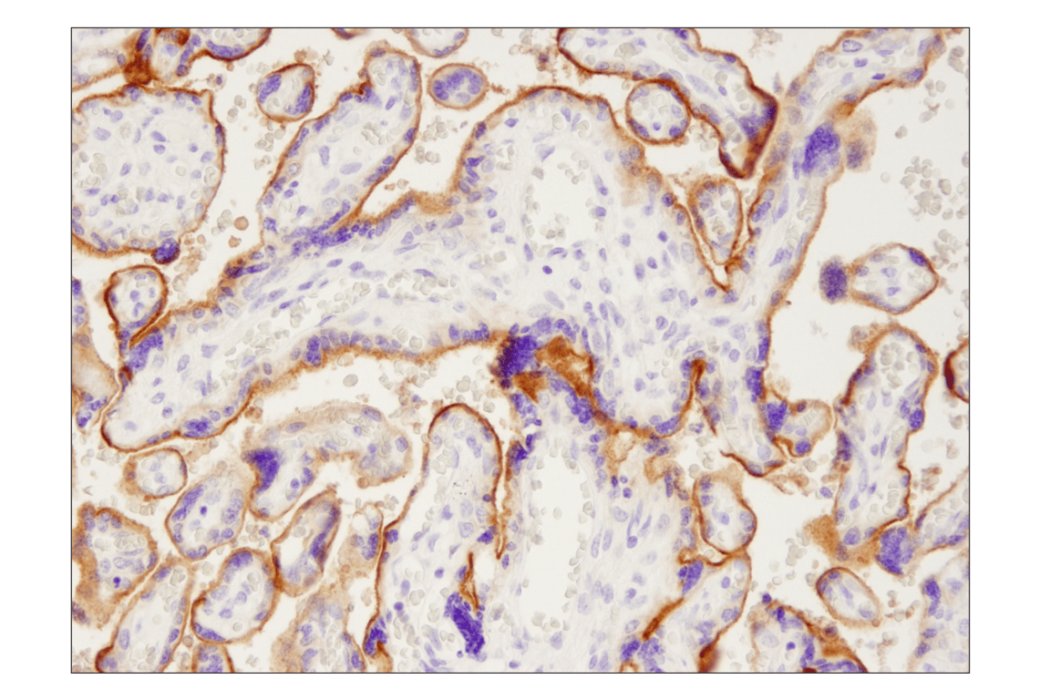
Immunohistochemical analysis of parffin-embedded human placenta using PD-L1 (405.9A11) Mouse mAb.