


 全部商品分类


BD Horizon™ BB700 Rat Anti-Mouse CD274


 下载产品说明书
 用小程序,查商品更便捷


 收藏
收藏
 对比
对比 咨询
咨询

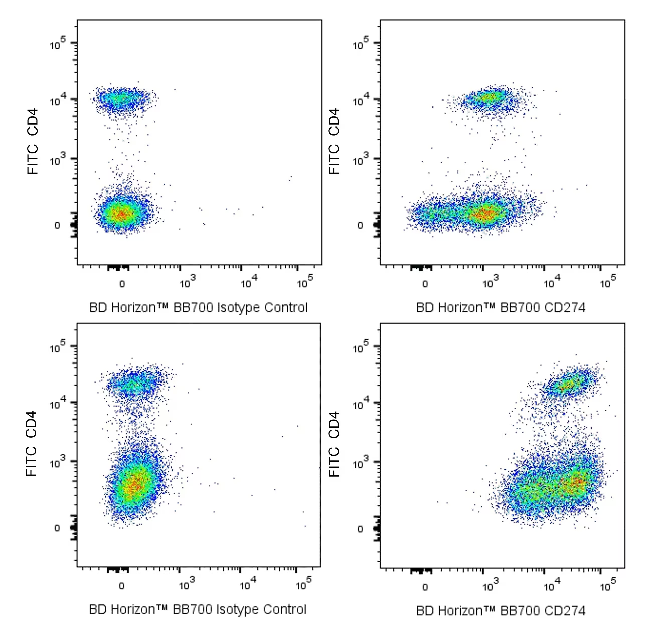
参考图片
Two-color flow cytometric analysis of CD274 expression on resting and activated mouse splenic leucocytes. Mouse splenic leucocytes were either not activated (Upper Plots) or activated (Lower Plots) by culture with plate-bound Purified NA/LE Hamster Anti-Mouse CD3 antibody (Cat. No. 553057) for 3 days (37°C). The cells were harvested, washed and preincubated with Purified Rat Anti-Mouse CD16/CD32 antibody (Mouse BD Fc Block™) (Cat. No. 553141/553142). The cells were then stained with FITC Rat Anti-Mouse CD4 antibody (Cat. No. 553729/557307/561828) and either BD Horizon™ BB700 Rat IgG2a, κ Isotype Control (Cat. No. 566413; Left Plots) or BD Horizon BB700 Rat Anti-Mouse CD274 antibody (Cat. No. 566879; Right Plots) at 0.25 µg/test. DAPI (4',6-Diamidino-2-Phenylindole, Dihydrochloride) Solution (Cat. No. 564907) was added to cells right before analysis. Bivariate pseudocolor density showing the correlated expression of CD274 (or Ig Isotype control staining) versus CD4 were derived from gated events with the forward and side light- scatter characteristics of viable (DAPI-negative) leucocytes. Flow cytometry and data analysis were performed using a BD LSRFortessa™ Cell Analyzer System and FlowJo™ software. Data shown on this Technical Data Sheet are not lot specific.