老带新
PD-L1 (Extracellular Domain Specific) (D8T4X) Rabbit mAb
1/2

PD-L1 (Extracellular Domain Specific) (D8T4X) Rabbit mAb

分享
品牌: CST
pdf 下载产品说明书
pdf 下载COA
pdf 下载SDS
用小程序,查商品更便捷
收藏
对比对比
咨询咨询
反应种属:
Human
来源宿主:
Rabbit
产品介绍
产品介绍
产品信息
荧光素标记
来源纯化

Monoclonal antibody is produced by immunizing animals with mammalian cells expressing full length PD-L1 protein.

宿主
Rabbit
商品描述

Product Usage Information

ApplicationDilution
Immunofluorescence (Immunocytochemistry)1:200
Flow Cytometry (Fixed/Permeabilized)1:50 - 1:200
Flow Cytometry (Live)1:50 - 1:200
同种型
Rabbit IgG
应用
目标/特异性

Specificity/Sensitivity

PD-L1 (Extracellular Domain Specific) (D8T4X) Rabbit mAb recognizes endogenous levels of total PD-L1 protein.

Species Reactivity:

Human

敏感性
Endogenous
背景
背景
Programmed cell death 1 ligand 1 (PD-L1, B7-H1, CD274) is a member of the B7 family of cell surface ligands that regulate T cell activation and immune responses. The PD-L1 ligand binds the PD-1 transmembrane receptor and inhibits T cell activation. PD-L1 was discovered following a search for novel B7 protein homologs and was later shown to be expressed by antigen presenting cells, activated T cells, and tissues including placenta, heart, and lung (1-3). Similar in structure to related B7 family members, PD-L1 protein contains extracellular IgV and IgC domains and a short, cytoplasmic region. Research studies demonstrate that PD-L1 is expressed in several tumor types, including melanoma, ovary, colon, lung, breast, and renal cell carcinomas (4-6). Expression of PD-L1 in cancer is associated with tumor-infiltrating lymphocytes, which mediate PD-L1 expression through the release of interferon gamma (7). Additional research links PD-L1 expression to cancers associated with viral infections (8,9). 1.Dong, H. et al. (1999) Nat Med 5, 1365-9. 2.Freeman, G.J. et al. (2000) J Exp Med 192, 1027-34. 3.Liang, S.C. et al. (2003) Eur J Immunol 33, 2706-16. 4.Dong, H. et al. (2002) Nat Med 8, 793-800. 5.Thompson, R.H. et al. (2006) Cancer Res 66, 3381-5. 6.Pardoll, D.M. (2012) Nat Rev Cancer 12, 252-64. 7.Taube, J.M. et al. (2012) Sci Transl Med 4, 127ra37. 8.Lyford-Pike, S. et al. (2013) Cancer Res 73, 1733-41. 9.Chen, B.J. et al. (2013) Clin Cancer Res 19, 3462-73.
研究领域
癌症,免疫学和肿瘤学,
翻译后修饰
unmodified
制备和贮存
保存方式

Supplied in 10 mM sodium HEPES (pH 7.5), 150 mM NaCl, 100 µg/ml BSA, 50% glycerol and less than 0.02% sodium azide. Store at –20°C. Do not aliquot the antibody.

For a carrier free (BSA and azide free) version of this product see product #58433.

数据库链接
Entrez-Gene ID
29126
UniProt ID
Q9NZQ7

参考图片

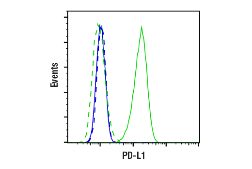
Flow cytometric analysis of live Ramos cells (blue, negative) and MDA-MB-231 cells (green, positive) using PD-L1 (Extracellular Domain Specific) (D8T4X) Rabbit mAb (solid lines) or concentration-matched Rabbit (DA1E) mAb IgG XP® Isotype Control #3900 (dashed lines). Anti-rabbit IgG (H+L), F(ab')2 Fragment (Alexa Fluor® 488 Conjugate) #4412 was used as a secondary antibody.

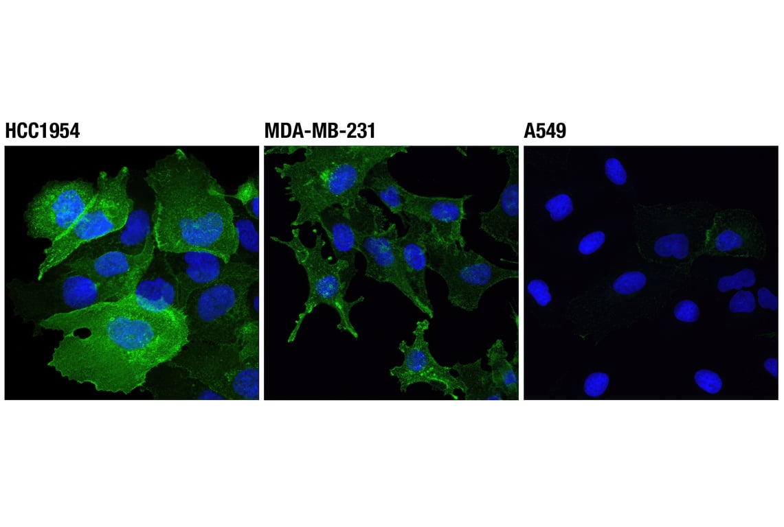
Confocal immunofluorescent analysis of HCC1954 (left), MDA-MB-231 (center), and A549 (right) cells using PD-L1 (Extracellular Domain Specific) (D8T4X) Rabbit mAb (green). Blue pseudocolor = DRAQ5® #4084 (fluorescent DNA dye).

声明 :本官网所有报价均为常温或者蓝冰运输价格,如有产品需要干冰运输,需另外加收干冰运输费。
货号:
86744S
一键复制
询价
供应商现货
20μl
100ul
选择数量
当前规格1件起购 
配送至
预计5-6个工作日送达,快递: 免运费,若需干冰额外收费