


 全部商品分类
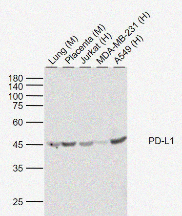


Rabbit anti-PD-L1 Monoclonal Antibody

 下载产品说明书
 用小程序,查商品更便捷


 收藏
收藏
 对比
对比 咨询
咨询Disclaimer note: The observed molecular weight of the protein may vary from the listed predicted molecular weight due to post translational modifications, post translation cleavages, relative charges, and other experimental factors.

Disclaimer note: The observed molecular weight of the protein may vary from the listed predicted molecular weight due to post translational modifications, post translation cleavages, relative charges, and other experimental factors.
参考图片
Sample: Lane 1: Lung (Mouse) Lysate at 40 ug Lane 2: Placenta (Mouse) Lysate at 40 ug Lane 3: Jurkat (Human) Cell Lysate at 30 ug Lane 4: MDA-MB-231 (Human) Cell Lysate at 30 ug Lane 5: A549 (Human) Cell Lysate at 30 ug Primary: Anti-PD-L1 at 1/1000 dilution Secondary: IRDye800CW Goat Anti-Rabbit IgG at 1/20000 dilution Predicted band size: 33 kDa Observed band size: 45 kDa
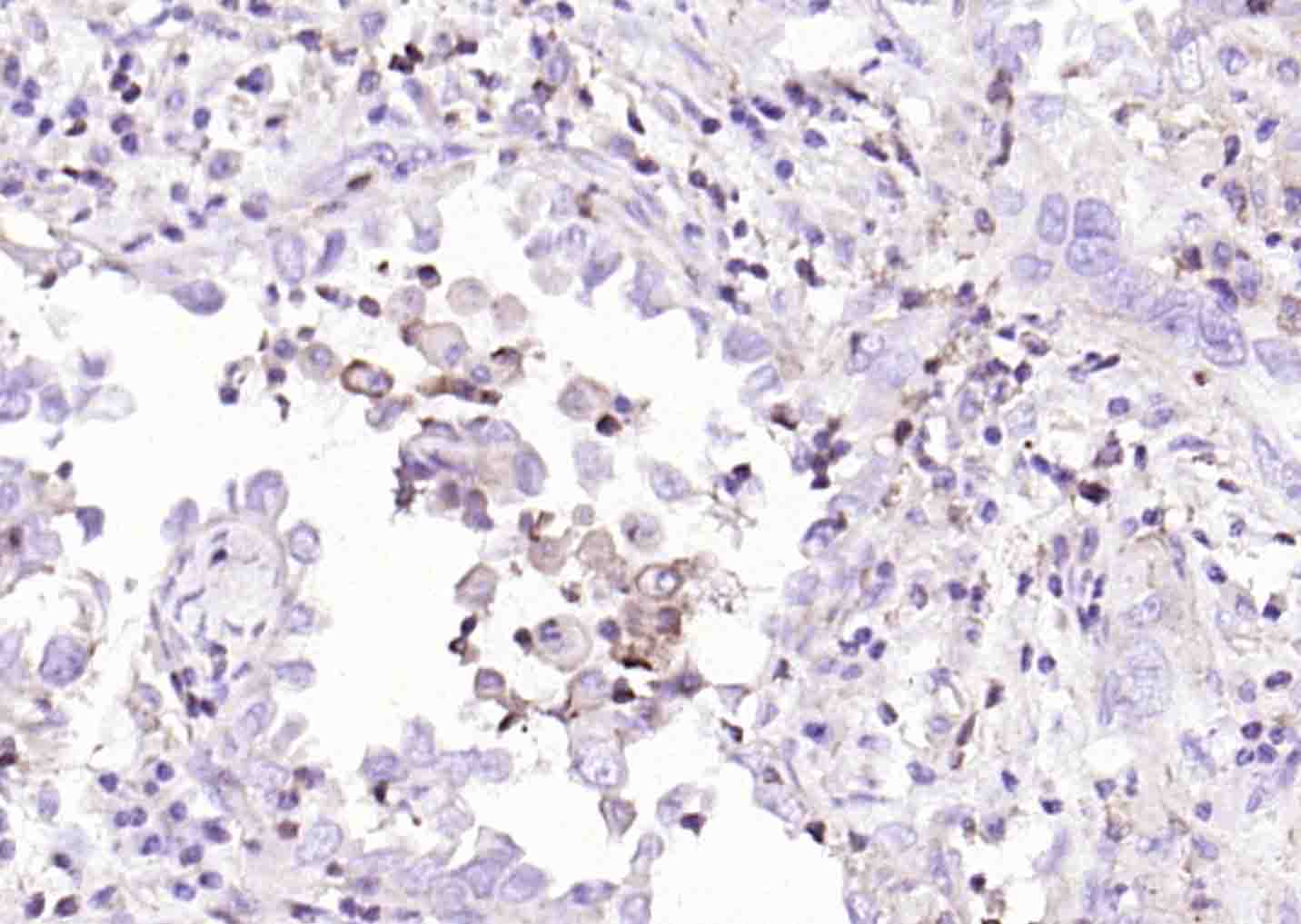
Paraformaldehyde-fixed, paraffin embedded (human lung carcinoma); Antigen retrieval by boiling in sodium citrate buffer (pH6.0) for 15min; Block endogenous peroxidase by 3% hydrogen peroxide for 20 minutes; Blocking buffer (normal goat serum) at 37°C for 30min; Incubation with (PD-L1) Monoclonal Antibody, Unconjugated at 1:200 overnight at 4°C, followed by operating according to SP Kit(Rabbit) instructionsand DAB staining.
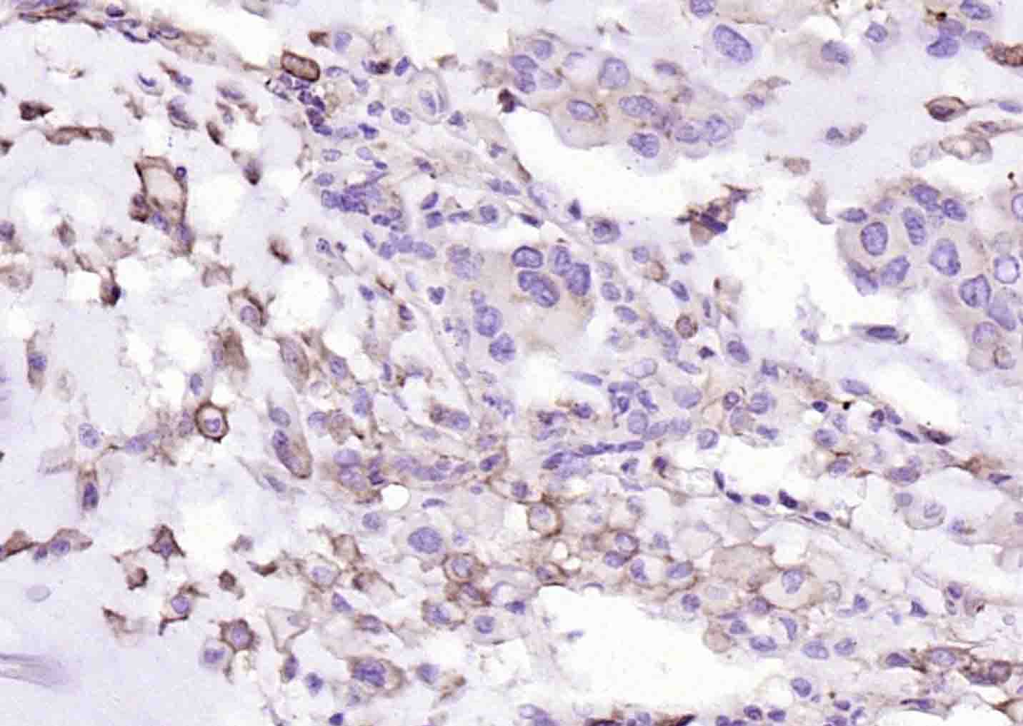
Paraformaldehyde-fixed, paraffin embedded (human cervical carcinoma); Antigen retrieval by boiling in sodium citrate buffer (pH6.0) for 15min; Block endogenous peroxidase by 3% hydrogen peroxide for 20 minutes; Blocking buffer (normal goat serum) at 37°C for 30min; Incubation with (PD-L1) Monoclonal Antibody, Unconjugated at 1:200 overnight at 4°C, followed by operating according to SP Kit(Rabbit) instructionsand DAB staining.