




 全部商品分类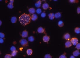


Human CD38 MAb (Clone 240742) (25 ug)


 用小程序,查商品更便捷



 收藏
收藏
 对比
对比 咨询
咨询Immunoprecipitation(25 µg/mL)
Immunocytochemistry(8-25 µg/mL)

Met1-Ile300
Accession # P28907
Human CD38 Antibody Summary
Met1-Ile300
Accession # P28907
*Small pack size (-SP) is supplied either lyophilized or as a 0.2 µm filtered solution in PBS.
Applications
Please Note: Optimal dilutions should be determined by each laboratory for each application. General Protocols are available in the Technical Information section on our website.
Scientific Data
 View Larger
CD38 in Human PBMCs. CD38 was detected in immersion fixed human peripheral blood mononuclear cells (PBMCs) using 10 µg/mL Mouse Anti-Human CD38 Monoclonal Antibody (Catalog # MAB2404) for 3 hours at room temperature. Cells were stained with the NorthernLights™ 557-conjugated Anti-Mouse IgG Secondary Antibody (red; NL007) and counterstained with DAPI (blue). View our protocol for Fluorescent ICC Staining of Non-adherent Cells.
 View Larger
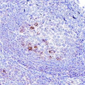
Detection of CD38 in human tonsil. CD38 was detected in immersion fixed paraffin-embedded sections of human tonsil using Mouse Anti-Human CD38 Monoclonal Antibody (Catalog # MAB2404) at 5 µg/mL for 1 hour at room temperature followed by incubation with the Anti-Mouse IgG VisUCyte™ HRP Polymer Antibody (Catalog # VC001). Before incubation with the primary antibody, tissue was subjected to heat-induced epitope retrieval using VisUCyte Antigen Retrieval Reagent-Basic (Catalog # VCTS021). Tissue was stained using DAB (brown) and counterstained with hematoxylin (blue). Specific staining was localized to cell surface in lymphocytes. View our protocol for IHC Staining with VisUCyte HRP Polymer Detection Reagents.
 View Larger
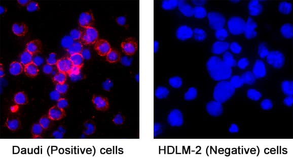
Detection of CD38 in Daudi Human Burkitt's Lymphoma Cell Line (positive) and HDLM‑2 Human Hodgkin’s Lymphoma Cell Line (negative) Cells. CD38 was detected in immersion fixed Daudi Human Burkitt's Lymphoma Cell Line (positive) and HDLM‑2 Human Hodgkin’s Lymphoma Cell Line (negative) Cells using Mouse Anti-Human CD38 Monoclonal Antibody (Catalog # MAB2404) at 5 µg/mL for 3 hours at room temperature. Cells were stained using the NorthernLights™ 557-conjugated Anti-Mouse IgG Secondary Antibody (red; Catalog # NL007) and counterstained with DAPI (blue). Specific staining was localized to cell surface and cytoplasm. View our protocol for Fluorescent ICC Staining of Non-adherent Cells.
 View Larger
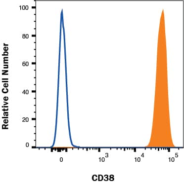
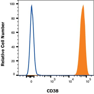
Detection of CD38 in Daudi cells by Flow Cytometry Daudi cells were stained with Mouse Anti-Human CD38 Monoclonal Antibody (Catalog # MAB2404, filled histogram) or isotype control antibody (Catalog # MAB003, open histogram) followed by Allophycocyanin-conjugated Anti-Mouse IgG Secondary Antibody (Catalog # F0101B). View our protocol for Staining Membrane-associated Proteins.
Reconstitution Calculator
Preparation and Storage
- 12 months from date of receipt, -20 to -70 °C as supplied.
- 1 month, 2 to 8 °C under sterile conditions after reconstitution.
- 6 months, -20 to -70 °C under sterile conditions after reconstitution.
Background: CD38
CD38, also known as ADP-ribosyl cyclase, is a Type II integral membrane protein. The enzyme is able to transform NAD(P)+ into three different products with calcium mobilizing ability, cyclic ADP-ribose, NAADP+, and ADP-ribose (1). CD38 is expressed in B and T lymphocytes, osteoclasts, and in cardiac, pancreatic, liver and kidney cells (2, 3). Through its production of cyclic ADP-ribose, CD38 modulates calcium-mediated signal transduction in many types of cells, including neutrophils and pancreatic beta cells (4, 5).
- Schuber, F. and F.E. Lund (2004) Curr. Mol. Med. 4:249.
- Jackson, D.G. and J.I. Bell (1990) J. Immunol. 144:2811.
- Sun, L. et al. (1999) J. Cell Biol. 146:1161.
- Partida-Sanchez, S. et al. (2001) Nature Med. 7:1209.
- Kato, I. et al. (1995) J. Biol. Chem. 270:30045.
Product Datasheets
Immunoprecipitation(25 µg/mL)
Immunocytochemistry(8-25 µg/mL)
Background: CD38
CD38, also known as ADP-ribosyl cyclase, is a Type II integral membrane protein. The enzyme is able to transform NAD(P)+ into three different products with calcium mobilizing ability, cyclic ADP-ribose, NAADP+, and ADP-ribose (1). CD38 is expressed in B and T lymphocytes, osteoclasts, and in cardiac, pancreatic, liver and kidney cells (2, 3). Through its production of cyclic ADP-ribose, CD38 modulates calcium-mediated signal transduction in many types of cells, including neutrophils and pancreatic beta cells (4, 5).
- Schuber, F. and F.E. Lund (2004) Curr. Mol. Med. 4:249.
- Jackson, D.G. and J.I. Bell (1990) J. Immunol. 144:2811.
- Sun, L. et al. (1999) J. Cell Biol. 146:1161.
- Partida-Sanchez, S. et al. (2001) Nature Med. 7:1209.
- Kato, I. et al. (1995) J. Biol. Chem. 270:30045.
Preparation and Storage
- 12 months from date of receipt, -20 to -70 °C as supplied.
- 1 month, 2 to 8 °C under sterile conditions after reconstitution.
- 6 months, -20 to -70 °C under sterile conditions after reconstitution.