Clone SP34-2 is a mouse IgG1 isotype monoclonal antibody, descendant of SP34 (mouse IgG3), with the same specificity and reactivity pattern as the parent clone. It cross-reacts with a major subset of peripheral blood lymphocytes, but not monocytes or granulocytes, of baboon, and rhesus, cynomolgus, and pigtail macaque monkeys. The distribution on lymphocytes is similar to that observed with normal human donor lymphocytes with the majority of CD3-positive cells being negative when dual stained with antibodies to B or NK cells markers. SP34-2 is also capable of inducing cell proliferation on both human and non-human primate PBMC.
商品描述
SP34-2
Clone SP34-2 is a mouse IgG1 isotype monoclonal antibody, descendant of SP34 (mouse IgG3), with the same specificity and reactivity pattern as the parent clone. It cross-reacts with a major subset of peripheral blood lymphocytes, but not monocytes or granulocytes, of baboon, and rhesus, cynomolgus, and pigtail macaque monkeys. The distribution on lymphocytes is similar to that observed with normal human donor lymphocytes with the majority of CD3-positive cells being negative when dual stained with antibodies to B or NK cells markers. SP34-2 is also capable of inducing cell proliferation on both human and non-human primate PBMC.
同种型
Mouse BALB/c IgG1, λ
克隆号
克隆 SP34-2 (RUO)
产品详情
PE
R-Phycoerythrin (PE), is part of the BD family of Phycobiliprotein dyes. This fluorochrome is a multimeric fluorescent phycobiliprotein with excitation maximum (Ex Max) of 496 nm and 566 nm and an emission maximum (Em Max) at 576 nm. PE is designed to be excited by the Blue (488 nm), Green (532 nm) and Yellow-Green (561 nm) lasers and detected using an optical filter centered near 575 nm (e.g., a 575/26-nm bandpass filter). As PE is excited by multiple lasers, this can result in cross-laser excitation and fluorescence spillover on instruments with various combinations of Blue, Green, and Yellow-Green lasers. Please ensure that your instrument’s configurations (lasers and optical filters) are appropriate for this dye.
PE
Yellow-Green 488 nm, 532 nm, 561 nm
496 nm, 566 nm
576 nm
应用
实验应用
Flow cytometry (Routinely Tested)
推荐用量
20 µl
反应种属
Rhesus, Cynomolgus, Baboon (QC Testing), Human (Tested in Development)
目标/特异性
CD3
背景
别名
CD3E; CD3-epsilon; T3E; TCRE
制备和贮存
存储溶液
Aqueous buffered solution containing BSA and ≤0.09% sodium azide.
保存方式
Aqueous buffered solution containing BSA and ≤0.09% sodium azide.
文献
文献
研发参考(6)
1. Blumberg RS, Ley S, Sancho J, et al. Structure of the T-cell antigen receptor: evidence for two CD3 epsilon subunits in the T-cell receptor-CD3 complex. Proc Natl Acad Sci U S A. 1990; 87(18):7220-7224. (Clone-specific).
2. Carter DL, Shieh TM, Blosser RL et al. CD56 identifies monocytes and not natural killer cells in rhesus macaques. Cytometry. 1999; 37(1):41-50. (Biology).
3. Pessano S, Oettgen H, Bhan AK, Terhorst C. The T3/T cell receptor complex: antigenic distinction between the two 20-kd T3 (T3-delta and T3-epsilon) subunits. EMBO J. 1985; 4(2):337-344. (Immunogen).
4. Sancho J, Ledbetter JA, Choi MS, Kanner SB, Deans JP, Terhorst C. CD3-zeta surface expression is required for CD4-p56lck-mediated upregulation of T cell antigen receptor-CD3 signaling in T cells. J Biol Chem. 1992; 267(11):7871-7879. (Biology).
5. Schlossman SF. Stuart F. Schlossman .. et al., ed. Leucocyte typing V : white cell differentiation antigens : proceedings of the fifth international workshop and conference held in Boston, USA, 3-7 November, 1993. Oxford: Oxford University Press; 1995.
6. Wilson AD, Shooshtari M, Finerty S, Watkins P, Morgan AJ. Selection of monoclonal antibodies for the identification of lymphocyte surface antigens in the New World primate Saguinus oedipus oedipus (cotton top tamarin). J Immunol Methods. 1995; 178(2):195-200. (Biology).
参考图片
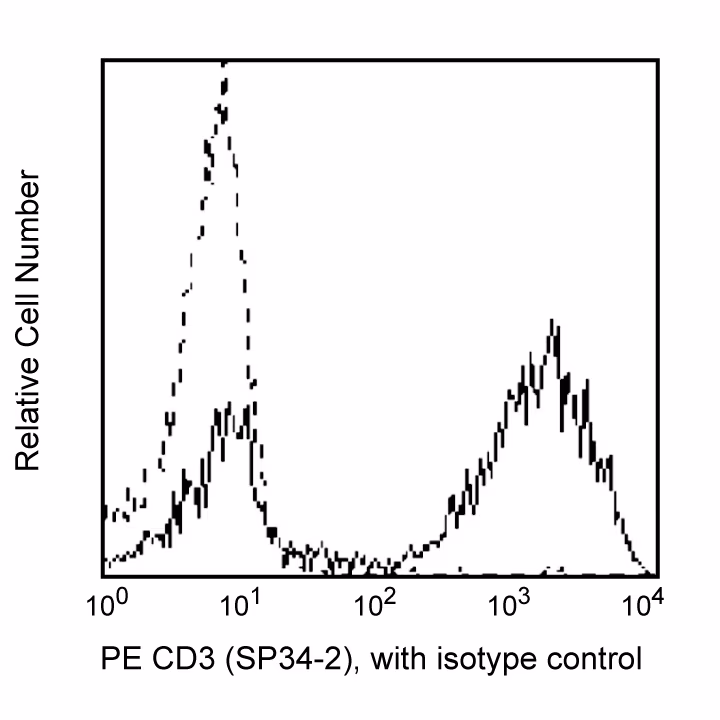
Flow cytometric analysis of CD3 expression on Rhesus macaque (Macaca mulatta) peripheral blood lymphocytes. Rhesus whole blood was stained with either PE Mouse Anti-Human CD3 (Cat. No. 552127; solid line histogram) or PE Mouse IgG1, κ Isotype Control (Cat. No. 556650; dashed line histogram). Erythrocytes were lysed with BD Pharm Lyse™ Lysing Buffer (Cat. No. 555899). Fluorescent histograms were derived from gated events with the side and forward light-scattering characteristics of viable lymphocytes.
Flow cytometric analysis of CD3 expression on Rhesus macaque (Macaca mulatta) peripheral blood lymphocytes. Rhesus whole blood was stained with either PE Mouse Anti-Human CD3 (Cat. No. 552127; solid line histogram) or PE Mouse IgG1, κ Isotype Control (Cat. No. 556650; dashed line histogram). Erythrocytes were lysed with BD Pharm Lyse™ Lysing Buffer (Cat. No. 555899). Fluorescent histograms were derived from gated events with the side and forward light-scattering characteristics of viable lymphocytes.


 全部商品分类


 下载产品说明书
 用小程序,查商品更便捷


 收藏
收藏
 对比
对比 咨询
咨询
