


 全部商品分类


1/2
BD Pharmingen™ FITC Mouse Anti-Human CD3


品牌: BD Pharmingen
 下载产品说明书 下载SDS
 用小程序,查商品更便捷


 收藏
收藏
 对比
对比 咨询
咨询反应种属:
Human (QC Testing)
来源宿主:
Mouse IgG2a, κ
产品介绍
产品使用步骤推荐 产品介绍
产品信息
荧光素标记
抗原名称
CD3

宿主
Mouse IgG2a, κ
简单描述
The HIT3a monoclonal antibody specifically binds to the human CD3ε-chain, a 20 kDa subunit of the CD3/T cell antigen receptor complex found on 70-80% of normal human peripheral blood lymphocytes and 60-85% of thymocytes. Studies from the HLDA Workshop show that this antibody can be mitogenic for T lymphocytes. The CD3 complex plays a role in signal transduction during antigen recognition by the T cell receptor. HIT3a antibody does not stain intracellular CD3 unlike the other CD3-specific clone, UCHT1 (Cat. No. 555330/550368).
商品描述
HIT3a
The HIT3a monoclonal antibody specifically binds to the human CD3ε-chain, a 20 kDa subunit of the CD3/T cell antigen receptor complex found on 70-80% of normal human peripheral blood lymphocytes and 60-85% of thymocytes. Studies from the HLDA Workshop show that this antibody can be mitogenic for T lymphocytes. The CD3 complex plays a role in signal transduction during antigen recognition by the T cell receptor. HIT3a antibody does not stain intracellular CD3 unlike the other CD3-specific clone, UCHT1 (Cat. No. 555330/550368).
同种型
Mouse IgG2a, κ
克隆号
克隆 HIT3a (RUO)
产品详情
FITC
Fluorescein (FITC) is part of the BD blue family of dyes. This is a small organic fluorochrome with an excitation maximum (Ex Max) at 494-nm and an emission maximum (Em Max) at 518-nm. FITC is designed to be excited by the Blue laser (488-nm) and detected using an optical filter centered near 520 nm (e.g., a 530/30-nm bandpass filter). Please ensure that your instrument’s configurations (lasers and optical filters) are appropriate for this dye.

FITC
Blue 488 nm
494 nm
518 nm
应用
实验应用
Flow cytometry (Routinely Tested)
推荐用量
20 µl
反应种属
Human (QC Testing)
目标/特异性
CD3
背景
别名
CD3E; T3E; TCRE; cd 3; cd-3; cd3; CD3-epsilon; 916
制备和贮存
存储溶液
Aqueous buffered solution containing BSA, protein stabilizer, and ≤0.09% sodium azide.
保存方式
Aqueous buffered solution containing BSA, protein stabilizer, and ≤0.09% sodium azide.
文献
文献
研发参考(6)
1. Barclay NA, Brown MH, Birkeland ML, et al, ed. The Leukocyte Antigen FactsBook. San Diego, CA: Academic Press; 1997.
2. Beverley PC, Callard RE. Distinctive functional characteristics of human "T" lymphocytes defined by E rosetting or a monoclonal anti-T cell antibody. Eur J Immunol. 1981; 11(4):329-334. (Biology).
3. Knapp W. W. Knapp .. et al., ed. Leucocyte typing IV : white cell differentiation antigens. Oxford New York: Oxford University Press; 1989:1-1182.
4. Lanier LL, Allison JP, Phillips JH. Correlation of cell surface antigen expression on human thymocytes by multi-color flow cytometric analysis: implications for differentiation. J Immunol. 1986; 137(8):2501-2507. (Biology).
5. McMichael AJ. A.J. McMichael .. et al., ed. Leucocyte typing III : white cell differentiation antigens. Oxford New York: Oxford University Press; 1987:1-1050.
6. Schlossman SF. Stuart F. Schlossman .. et al., ed. Leucocyte typing V : white cell differentiation antigens : proceedings of the fifth international workshop and conference held in Boston, USA, 3-7 November, 1993. Oxford: Oxford University Press; 1995.
参考图片
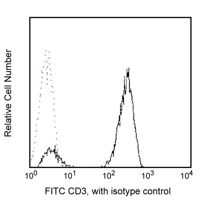
Flow cytometric analysis of CD3 expression on human peripheral blood lymphocytes. Whole blood was stained with either FITC Mouse IgG2a, κ Isotype Control (Cat. No. 555573; dashed line histogram) or FITC Mouse Anti-Human CD3 (Cat. No. 561802/555539; solid line histogram). Erythrocytes were lysed with Lysing Buffer (Cat. No. 555899). The fluorescence histograms showing CD3 expression (or Ig Isotype control staining) were derived from gated events with the forward and side light-scattering characteristics of viable lymphocytes. Flow cytometry was performed on a BD FACScan™.
声明 :本官网所有报价均为常温或者蓝冰运输价格,如有产品需要干冰运输,需另外加收干冰运输费。