


 全部商品分类


BD Pharmingen™ APC-Cy™7 Mouse Anti-Human CD3

 下载产品说明书 下载SDS
 用小程序,查商品更便捷


 收藏
收藏
 对比
对比 咨询
咨询
参考图片
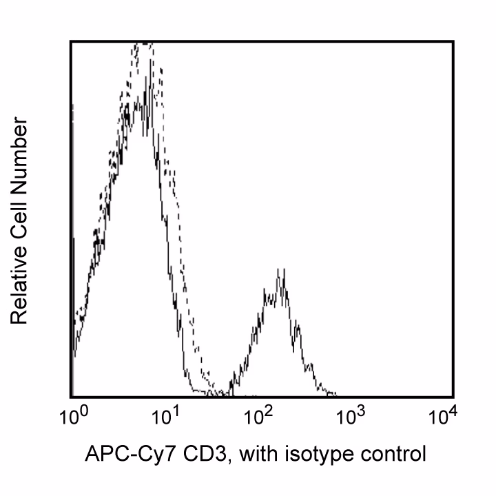
Flow cytometric analysis of CD3 expression on Rhesus macaque (Macaca mulatta) peripheral blood lymphocytes. Whole rhesus blood was stained with either APC-Cy™7 Mouse IgG1, κ Isotype Control (Cat. No. 557873; dashed line histogram) or APC-Cy™7 Mouse Anti-Human CD3 (Cat. No. 557757; solid line histogram). Erythrocytes were lysed with BD FACS™ Lysing Solution (Cat. No. 349202). Fluorescent histograms were derived from gated events with the side and forward light-scatter characteristics of viable lymphocytes.
Flow cytometric analysis of CD3 expression on Rhesus macaque (Macaca mulatta) peripheral blood lymphocytes. Whole rhesus blood was stained with either APC-Cy™7 Mouse IgG1, κ Isotype Control (Cat. No. 557873; dashed line histogram) or APC-Cy™7 Mouse Anti-Human CD3 (Cat. No. 557757; solid line histogram). Erythrocytes were lysed with BD FACS™ Lysing Solution (Cat. No. 349202). Fluorescent histograms were derived from gated events with the side and forward light-scatter characteristics of viable lymphocytes.