Rat SD, also known as Sprague-Dawley (outbred) IgG2b, κ
免疫原
γδ TCR-positive T-T hybridoma D1
简单描述
The 17A2 monoclonal antibody specifically binds to the T-cell receptor-associated CD3 complex that is expressed on many thymocytes and mature T lymphocytes. Plate-bound 17A2 antibody has been reported to induce IL-2 production by cultured T cells in the absence of accessory cells. The binding of the 17A2 antibody to T cells can be blocked by the anti-CD3e mAb 145-2C11 (Cat. No. 557306/553058/550275). This suggests that the 17A2 antibody recognizes an epitope of the CD3 epsilon chain. In vivo treatment with 17A2 antibody has been reported to partially deplete T lymphocytes and temporarily down-modulates CD3 expression on T cells.
商品描述
17A2
The 17A2 monoclonal antibody specifically binds to the T-cell receptor-associated CD3 complex that is expressed on many thymocytes and mature T lymphocytes. Plate-bound 17A2 antibody has been reported to induce IL-2 production by cultured T cells in the absence of accessory cells. The binding of the 17A2 antibody to T cells can be blocked by the anti-CD3e mAb 145-2C11 (Cat. No. 557306/553058/550275). This suggests that the 17A2 antibody recognizes an epitope of the CD3 epsilon chain. In vivo treatment with 17A2 antibody has been reported to partially deplete T lymphocytes and temporarily down-modulates CD3 expression on T cells.
同种型
Rat SD, also known as Sprague-Dawley (outbred) IgG2b, κ
克隆号
克隆 17A2 (RUO)
浓度
0.2 mg/ml
产品详情
PerCP-Cy5.5
PerCP-Cy5.5 dye is part of the BD blue family of dyes. This tandem fluorochrome is comprised of a fluorescent protein complex (PerCP) with an excitation maximum (Ex Max) of 482 nm and an acceptor dye with an emission maximum (Em Max) at 676 nm. PerCP-Cy5 is designed to be excited by the blue laser (488-nm) and detected using an optical filter centered near 680 nm (e.g., a 695/40 nm bandpass filter). The donor dye can be partially excited by the Violet (405-nm) laser resulting in cross-laser excitation and fluorescence spillover. Please ensure that your instrument’s configurations (lasers and optical filters) are appropriate for this dye.
研发参考(3)
1. Miescher GC, Schreyer M, MacDonald HR. Production and characterization of a rat monoclonal antibody against the murine CD3 molecular complex. Immunol Lett. 1989; 23(2):113-118. (Immunogen).
2. Mysliwietz J, Thierfelder S. Antilymphocytic antibodies and marrow transplantation. XII. Suppression of graft-versus-host disease by T-cell-modulating and depleting antimouse CD3 antibody is most effective when preinjected in the marrow recipient. Blood. 1992; 80(10):2661-2667. (Biology).
3. Wu L, Antica M, Johnson GR, Scollay R, Shortman K. Developmental potential of the earliest precursor cells from the adult mouse thymus. J Exp Med. 1991; 174(6):1617-1627. (Biology).
参考图片
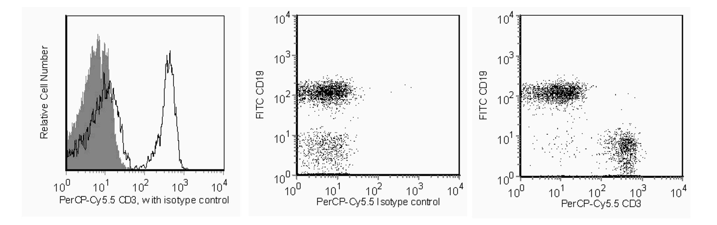
Flow cytometric analysis of CD3 on mouse splenocytes. Left Panel: Splenocytes from C57BL/6 mice were stained with either a PerCP-Cy™5.5 Rat IgG2b, κ isotype control (shaded) or with the PerCP-Cy™5.5 Rat Anti-Mouse CD3 antibody (unshaded). Middle and Right Panels: Splenocytes from C57BL/6 mice were stained with a FITC Rat Anti-Mouse CD19 antibody (Cat.No. 553785) in conjunction with either a PerCP-Cy™5.5 Rat IgG2b, κ isotype control (middle panel) or the PerCP-Cy™5.5 Rat Anti-Mouse CD3 antibody (right panel). Histograms and dot plots were derived from gated events based on light scattering characteristics for splenocytes. Flow cytometry was performed on a BD™ LSR II flow cytometry system.
Flow cytometric analysis of CD3 on mouse splenocytes. Left Panel: Splenocytes from C57BL/6 mice were stained with either a PerCP-Cy™5.5 Rat IgG2b, κ isotype control (shaded) or with the PerCP-Cy™5.5 Rat Anti-Mouse CD3 antibody (unshaded). Middle and Right Panels: Splenocytes from C57BL/6 mice were stained with a FITC Rat Anti-Mouse CD19 antibody (Cat.No. 553785) in conjunction with either a PerCP-Cy™5.5 Rat IgG2b, κ isotype control (middle panel) or the PerCP-Cy™5.5 Rat Anti-Mouse CD3 antibody (right panel). Histograms and dot plots were derived from gated events based on light scattering characteristics for splenocytes. Flow cytometry was performed on a BD™ LSR II flow cytometry system.


 全部商品分类


 下载产品说明书
 用小程序,查商品更便捷


 收藏
收藏
 对比
对比 咨询
咨询
