Human infant thymocytes and peripheral blood lymphocytes from a Sézary Syndrome donor
简单描述
The UCHT1 monoclonal antibody specifically binds to the human CD3ε-chain, a 20-kDa subunit of the CD3/T cell antigen receptor complex. CD3ε is expressed on 70-80% of normal human peripheral blood lymphocytes and 60-85% of thymocytes. Studies from the HLDA Workshop show that this antibody is mitogenic for CD3ε-positive cells when used in conjunction with costimulatory agents such as pokeweed mitogen or anti-CD28 antibody. CD3 plays a central role in signal transduction during antigen recognition. The UCHT1 antibody stains both surface and intracellular CD3ε unlike the other CD3 clone, HIT3a, that stains only extracellular CD3ε.
The antibody was conjugated to BD Horizon™ BV421 which is part of the BD Horizon Brilliant™ Violet family of dyes. With an Ex Max of 407-nm and Em Max at 421-nm, BD Horizon™ BV421 can be excited by the violet laser and detected in the standard Pacific Blue™ filter set (eg, 450/50-nm filter). BD Horizon™ BV421 conjugates are very bright, often exhibiting a 10 fold improvement in brightness compared to Pacific Blue™ conjugates.
商品描述
UCHT1
The UCHT1 monoclonal antibody specifically binds to the human CD3ε-chain, a 20-kDa subunit of the CD3/T cell antigen receptor complex. CD3ε is expressed on 70-80% of normal human peripheral blood lymphocytes and 60-85% of thymocytes. Studies from the HLDA Workshop show that this antibody is mitogenic for CD3ε-positive cells when used in conjunction with costimulatory agents such as pokeweed mitogen or anti-CD28 antibody. CD3 plays a central role in signal transduction during antigen recognition. The UCHT1 antibody stains both surface and intracellular CD3ε unlike the other CD3 clone, HIT3a, that stains only extracellular CD3ε.
The antibody was conjugated to BD Horizon™ BV421 which is part of the BD Horizon Brilliant™ Violet family of dyes. With an Ex Max of 407-nm and Em Max at 421-nm, BD Horizon™ BV421 can be excited by the violet laser and detected in the standard Pacific Blue™ filter set (eg, 450/50-nm filter). BD Horizon™ BV421 conjugates are very bright, often exhibiting a 10 fold improvement in brightness compared to Pacific Blue™ conjugates.
同种型
Mouse BALB/c IgG1, κ
克隆号
克隆 UCHT1 (also known as UCHT-1; UCHT 1) (RUO)
产品详情
BV421
The BD Horizon Brilliant Violet™ 421 (BV421) Dye is part of the BD Horizon Brilliant Violet™ family of dyes. This polymer-technology based dye has an excitation maximum (Ex Max) of 407-nm and an emission maximum (Em Max) at 423-nm. Driven by BD innovation, BV421 is designed to be excited by the violet laser (405-nm) and detected using an optical filter centered near 420-nm (e.g., a 431/28-nm or 450/50-nm bandpass filter). BV421 is an ideal alternative for V450 as it is approximately ten times brighter with less spillover into the BV510/V500 detector. Please ensure that your instrument’s configurations (lasers and optical filters) are appropriate for this dye.
BV421
Violet 405 nm
407 nm
423 nm
应用
实验应用
Flow cytometry (Routinely Tested), Immunofluorescence (Tested During Development)
Aqueous buffered solution containing BSA and ≤0.09% sodium azide.
保存方式
Aqueous buffered solution containing BSA and ≤0.09% sodium azide.
文献
文献
研发参考(6)
1. Beverley PC, Callard RE. Distinctive functional characteristics of human "T" lymphocytes defined by E rosetting or a monoclonal anti-T cell antibody. Eur J Immunol. 1981; 11(4):329-334. (Clone-specific).
2. Knapp W. W. Knapp .. et al., ed. Leucocyte typing IV : white cell differentiation antigens. Oxford New York: Oxford University Press; 1989:1-1182.
3. Lanier LL, Allison JP, Phillips JH. Correlation of cell surface antigen expression on human thymocytes by multi-color flow cytometric analysis: implications for differentiation. J Immunol. 1986; 137(8):2501-2507. (Biology).
4. McMichael AJ. A.J. McMichael .. et al., ed. Leucocyte typing III : white cell differentiation antigens. Oxford New York: Oxford University Press; 1987:1-1050.
5. Schlossman SF. Stuart F. Schlossman .. et al., ed. Leucocyte typing V : white cell differentiation antigens : proceedings of the fifth international workshop and conference held in Boston, USA, 3-7 November, 1993. Oxford: Oxford University Press; 1995.
6. Zola H. Leukocyte and stromal cell molecules : the CD markers. Hoboken, N.J.: Wiley-Liss; 2007.
参考图片
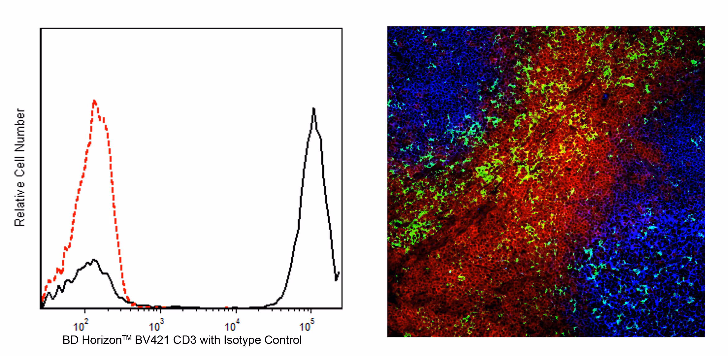
Flow cytometric analysis of CD3 expression on human peripheral blood lymphocytes (left panel). Human whole blood was stained with the BD Horizon™ BV421 Mouse anti-Human CD3 antibody (Cat. No. 562426/562427; solid line histogram) or with BD Horizon™ BV421 Mouse IgG1, κ Isotype Control (Cat. No. 562438; dashed line histogram). The erythrocytes were lysed with BD Pharm Lyse™ Lysing Buffer (Cat. No. 555899). The fluorescence histograms were derived from events with the forward and side light-scatter characteristics of viable lymphocytes. Flow cytometry was performed using a BD FACSCanto™ II Flow Cytometer System.
Immunohistofluorescent analysis of CD3 expression by cells within human tonsil (right panel). A human tonsil cryosection (5 µm) was fixed with BD Cytofix™ Fixation Buffer (Cat. No. 554655), blocked with 5% goat serum and 1% BSA diluted in 1x PBS, and stained with BD Pharmingen™ Purified Mouse Anti-Human CD11c antibody (Cat. No. 565805) followed by BD Horizon™ BV480 Goat Anti-Mouse Ig second step antibody (Cat. No. 564877, pseudo-colored green). Sections were thoroughly washed, then stained with BD Horizon™ BV421 Mouse Anti-Human CD3 antibody (Cat. No. 562426/562427, pseudo-colored red) and Alexa Fluor® 488 Mouse Anti-Human CD19 antibody (Cat. No. 557697, pseudo-colored blue). Images were captured on a standard epifluorescence microscope. Original magnification, 20x.
Flow cytometric analysis of CD3 expression on human peripheral blood lymphocytes (left panel). Human whole blood was stained with the BD Horizon™ BV421 Mouse anti-Human CD3 antibody (Cat. No. 562426/562427; solid line histogram) or with BD Horizon™ BV421 Mouse IgG1, κ Isotype Control (Cat. No. 562438; dashed line histogram). The erythrocytes were lysed with BD Pharm Lyse™ Lysing Buffer (Cat. No. 555899). The fluorescence histograms were derived from events with the forward and side light-scatter characteristics of viable lymphocytes. Flow cytometry was performed using a BD FACSCanto™ II Flow Cytometer System. Immunohistofluorescent analysis of CD3 expression by cells within human tonsil (right panel). A human tonsil cryosection (5 µm) was fixed with BD Cytofix™ Fixation Buffer (Cat. No. 554655), blocked with 5% goat serum and 1% BSA diluted in 1x PBS, and stained with BD Pharmingen™ Purified Mouse Anti-Human CD11c antibody (Cat. No. 565805) followed by BD Horizon™ BV480 Goat Anti-Mouse Ig second step antibody (Cat. No. 564877, pseudo-colored green). Sections were thoroughly washed, then stained with BD Horizon™ BV421 Mouse Anti-Human CD3 antibody (Cat. No. 562426/562427, pseudo-colored red) and Alexa Fluor® 488 Mouse Anti-Human CD19 antibody (Cat. No. 557697, pseudo-colored blue). Images were captured on a standard epifluorescence microscope. Original magnification, 20x.


 全部商品分类


 下载产品说明书
 用小程序,查商品更便捷


 收藏
收藏
 对比
对比 咨询
咨询
