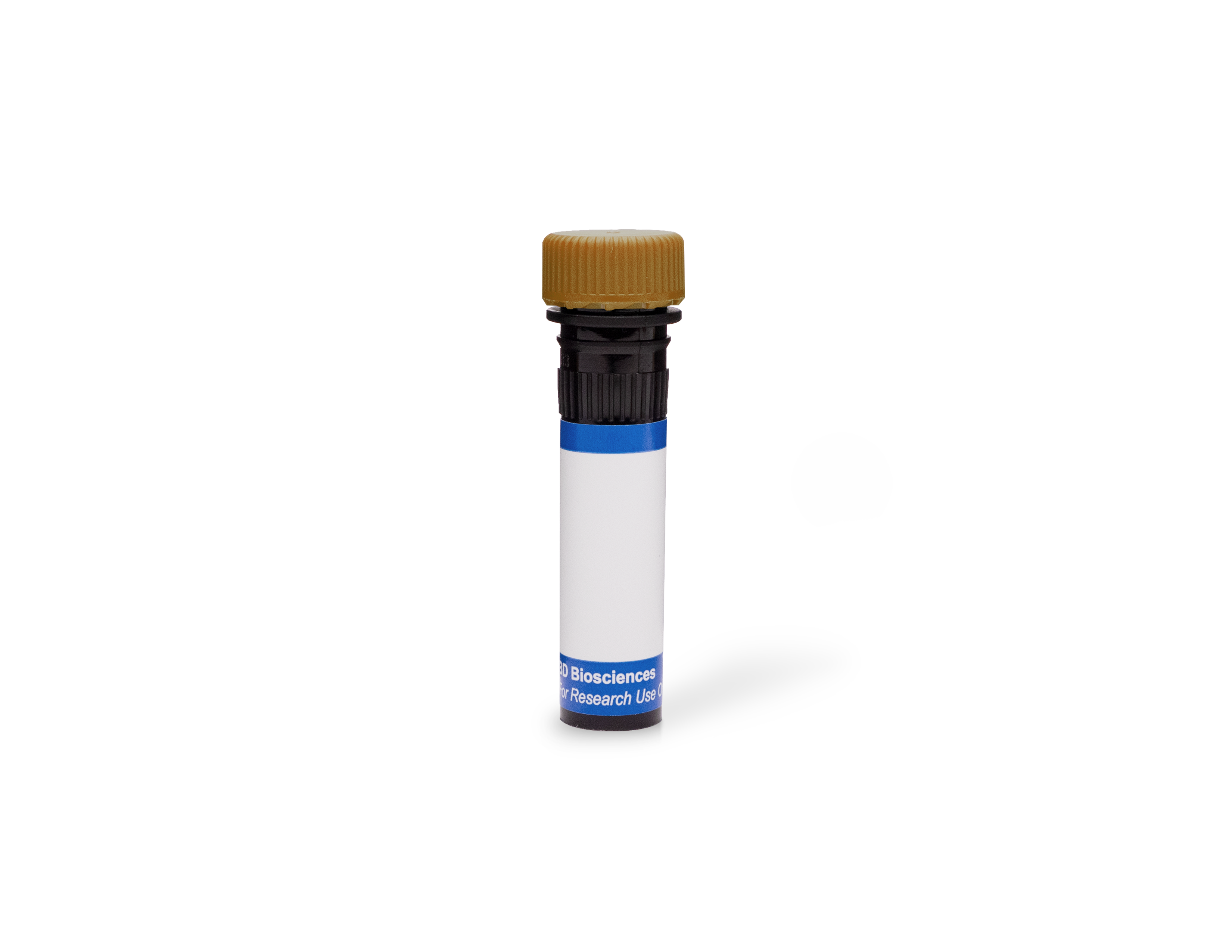
 全部商品分类


1/2

品牌: BD Pharmingen
 下载产品说明书
 下载SDS
下载SDS 用小程序,查商品更便捷


 收藏
收藏
 对比
对比 咨询
咨询反应种属:
Human (QC Testing)
来源宿主:
Mouse BALB/c IgG1, κ
产品介绍
产品使用步骤推荐 产品介绍
产品信息
荧光素标记
抗原名称
CD3

宿主
Mouse BALB/c IgG1, κ

免疫原
Human Thymocytes

简单描述
The SK7 (Leu-4) monoclonal antibody specifically binds to the epsilon chain of the CD3 antigen/T-cell antigen receptor (TCR) complex. This complex is composed of at least six proteins that range in molecular weight from 20 to 30 kDa. The antigen recognized by CD3 antibodies is noncovalently associated with either α/β or γ/δ TCR (70 to 90 kDa). The CD3 antigen is present on 61% to 85% of normal peripheral blood lymphocytes 60% to 85% of thymocytes and on Purkinje cells in the cerebellum. The soluble form of this antibody has a mitogenic effect on most peripheral blood T lymphocytes, provided appropriate functional monocytes are present.
The antibody was conjugated to BD Horizon BB700, which is part of the BD Horizon Brilliant™ Blue family of dyes. It is a polymer-based tandem dye developed exclusively by BD Biosciences. With an excitation max of 485 nm and an emission max of 693 nm, BD Horizon BB700 can be excited by the 488 nm laser and detected in a standard PerCP-Cy™5.5 set (eg, 695/40-nm filter). This dye provides a much brighter alternative to PerCP-Cy5.5 with less cross laser excitation off the 405 nm and 355 nm lasers.

商品描述
SK7
The SK7 (Leu-4) monoclonal antibody specifically binds to the epsilon chain of the CD3 antigen/T-cell antigen receptor (TCR) complex. This complex is composed of at least six proteins that range in molecular weight from 20 to 30 kDa. The antigen recognized by CD3 antibodies is noncovalently associated with either α/β or γ/δ TCR (70 to 90 kDa). The CD3 antigen is present on 61% to 85% of normal peripheral blood lymphocytes 60% to 85% of thymocytes and on Purkinje cells in the cerebellum. The soluble form of this antibody has a mitogenic effect on most peripheral blood T lymphocytes, provided appropriate functional monocytes are present.
The antibody was conjugated to BD Horizon BB700, which is part of the BD Horizon Brilliant™ Blue family of dyes. It is a polymer-based tandem dye developed exclusively by BD Biosciences. With an excitation max of 485 nm and an emission max of 693 nm, BD Horizon BB700 can be excited by the 488 nm laser and detected in a standard PerCP-Cy™5.5 set (eg, 695/40-nm filter). This dye provides a much brighter alternative to PerCP-Cy5.5 with less cross laser excitation off the 405 nm and 355 nm lasers.

同种型
Mouse BALB/c IgG1, κ

克隆号
克隆 SK7 (also known as Leu-4) (RUO)

产品详情
BB700
The BD Horizon Brilliant™ Blue 700 (BB700) Dye is part of the BD Horizon Brilliant™ Blue family of dyes. This tandem fluorochrome is comprised of a polymer-technology dye donor with an excitation maximum (Ex Max) of 476-nm and an acceptor dye with an emission maximum (Em Max) at 695-nm. Driven by BD innovation, BB700 is designed to be excited by the blue laser (488-nm) and detected using an optical filter centered near 695-nm (e.g., a 695/20-nm bandpass filter). The donor dye can be excited by the Violet (405 nm) laser and the acceptor dye can be excited by the red (627–640 nm) laser resulting in cross-laser excitation and fluorescence spillover. BB700 Reagents are significantly brighter than equivalent PerCP or PerCP-Cy5.5 reagents and are less sensitive to photobleaching. In addition, BB700 shows much less excitation by the violet (407-nm) laser resulting in less spillover. BB700 has minimal yellow green (562-nm) excitation and is ideal for instruments with both blue (488-nm) and yellow green (562-nm) lasers. Please ensure that your instrument’s configurations (lasers and optical filters) are appropriate for this dye.

BB700
Blue 488 nm
476 nm
695 nm
应用
实验应用
Flow cytometry (Routinely Tested)

推荐用量
5 µl

反应种属
Human (QC Testing)

目标/特异性
CD3

背景
别名
CD3-epsilon; CD3E; Leu4; T-cell surface antigen T3/Leu-4 epsilon chain; T3E

制备和贮存
存储溶液
Aqueous buffered solution containing ≤0.09% sodium azide.

保存方式
Aqueous buffered solution containing ≤0.09% sodium azide.
文献
文献
研发参考(13)
1. Ernst DN, Shih CC. CD3 complex. J Biol Regul Homeost Agents. 2000; 14(3):226-229. (Biology).
2. Kan EA, Wang CY, Wang LC, Evans RL. Noncovalently bonded subunits of 22 and 28 kd are rapidly internalized by T cells reacted with anti-Leu-4 antibody. J Immunol. 1983; 131(2):536-539. (Clone-specific: Flow cytometry, Functional assay, Immunofluorescence, Immunoprecipitation).
3. Kaneoka H, Perez-Rojas G, Sasasuki T, Benike CJ, Engleman EG. Human T lymphocyte proliferation induced by a pan-T monoclonal antibody (anti-Leu 4): heterogeneity of response is a function of monocytes. J Immunol. 1983; 131(1):158-164. (Clone-specific: Activation, Functional assay, Stimulation).
4. Knapp W. W. Knapp .. et al., ed. Leucocyte typing IV : white cell differentiation antigens. Oxford New York: Oxford University Press; 1989:1-1182.
5. Knowles RW. Immunochemical analysis of the T-cell–specific antigens. In: Reinherz EL. Ellis L. Reinherz .. et al., ed. Leukocyte typing II. New York: Springer-Verlag; 1986:259-288.
6. Kurrle R, Seyfert W, Trautwein A, Seiler FR. T cell activation by CD3 antibodies. In: Reinherz EL. Ellis L. Reinherz .. et al., ed. Leukocyte typing II. New York: Springer-Verlag; 1986:137-146.
7. Lanier LL, Allison JP, Phillips JH. Correlation of cell surface antigen expression on human thymocytes by multi-color flow cytometric analysis: implications for differentiation. J Immunol. 1986; 137(8):2501-2507. (Clone-specific: Immunoprecipitation).
8. Ledbetter JA, Evans RL, Lipinski M, Cunningham-Rundles C, Good RA, Herzenberg LA. Evolutionary conservation of surface molecules that distinguish T lymphocyte helper/inducer and cytotoxic/suppressor subpopulations in mouse and man. J Exp Med. 1981; 153(2):310-323. (Clone-specific: Immunoprecipitation).
9. Ledbetter JA, Frankel AE, Herzenberg. Human Leu T-cell differentiation antigens: quantitative expression on normal lymphoid cells and cell lines. In: Hammerling G, Hammerling U, Kearney J, ed. Monoclonal Antibodies and T Cell Hybridomas: Perspectives and Technical News. New York: Elsevier/North Holland Biomedical Press; 1981:16-22.
10. McMichael AJ. A.J. McMichael .. et al., ed. Leucocyte typing III : white cell differentiation antigens. Oxford New York: Oxford University Press; 1987:1-1050.
11. Schlossman SF. Stuart F. Schlossman .. et al., ed. Leucocyte typing V : white cell differentiation antigens : proceedings of the fifth international workshop and conference held in Boston, USA, 3-7 November, 1993. Oxford: Oxford University Press; 1995.
12. Zola H. Leukocyte and stromal cell molecules : the CD markers. Hoboken, N.J.: Wiley-Liss; 2007.
13. van Dongen JJM, Krissansen GW, Wolvers-Tettero ILM, et al. Cytoplasmic expression of the CD3 antigen as a diagnostic marker for immature T-cell malignanacies. Blood. 1988; 71(3):603-612. (Clone-specific: Immunofluorescence, Western blot).

数据库链接
Entrez-Gene ID
916

参考图片
Flow cytometric analysis of CD3 expression on human peripheral blood lymphocytes. Whole blood was stained with either BD Horizon™ BB700 Mouse IgG1, κ Isotype Control (Cat. No. 566404; dashed line histogram) or BD Horizon BB700 Mouse Anti-Human CD3 antibody (Cat. No. 566575/566576; solid line histogram). Erythrocytes were lysed with BD FACS™ Lysing Solution (Cat. No. 349202). The fluorescence histogram showing CD3 expression (or Ig Isotype control staining) was derived from gated events with the forward and side light-scatter characteristics of intact lymphocytes. Flow cytometric analysis was performed using a BD FACSCelesta™ Flow Cytometer System.
声明 :本官网所有报价均为常温或者蓝冰运输价格,如有产品需要干冰运输,需另外加收干冰运输费。


