


 全部商品分类


BD Pharmingen™ Purifed NA/LE Mouse Anti-Human CD3

 下载产品说明书 下载SDS
 用小程序,查商品更便捷


 收藏
收藏
 对比
对比 咨询
咨询
参考图片
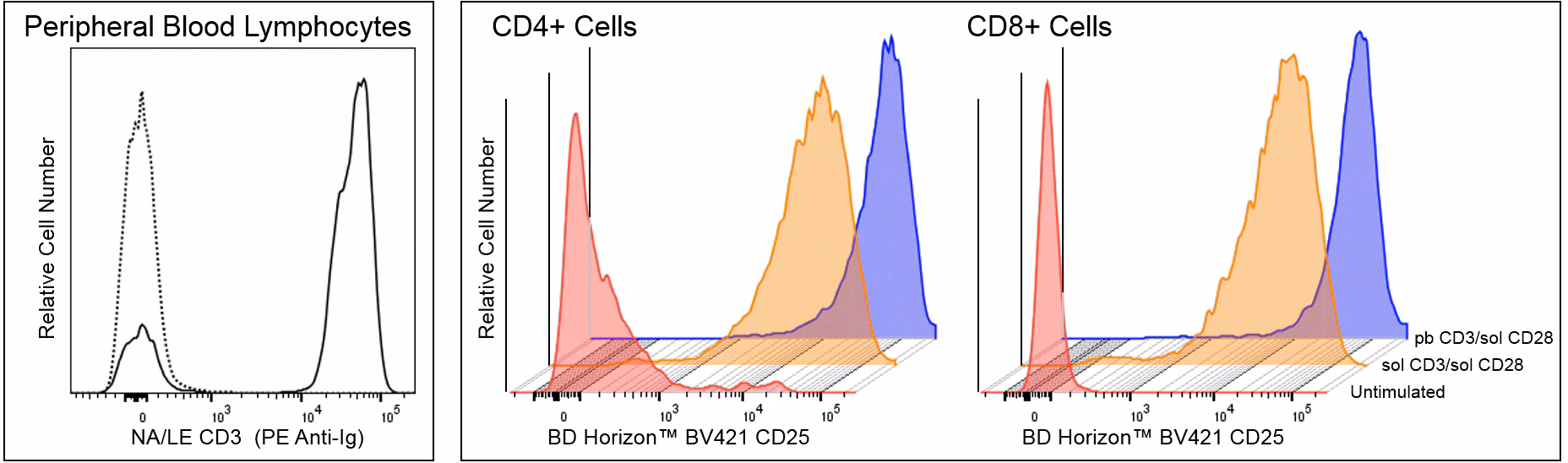
CD3 expression and function Left Panel -Flow cytometric analysis of CD3 expression on human peripheral blood lymphocytes (PBL). Peripheral blood was incubated with either Purified Mouse IgG2a κ Isotype Control (Cat. No. 553454; dashed line histogram) or Purified NA/LE Mouse Anti-Human CD3 antibody (Cat. No. 566685; solid line histogram). The cells were washed and stained with PE Goat Anti-Mouse Ig (Multiple Adsorption) (Cat. No. 550589). Erythrocytes were lysed with BD Pharm Lyse™ Lysing Buffer (Cat. No. 555899). The fluorescence histogram showing CD3 expression (or Ig Isotype control staining) was derived from gated events with the forward and side light-scatter characteristics of viable lymphocytes. Right Panel - Multicolor flow cytometric analysis of CD25 expression on unstimulated and Anti-CD3 plus Anti-CD28 antibody-stimulated human PBL. Human peripheral blood mononuclear cells (PBMC) were not stimulated [Unstimulated: Bottom red-colored histograms] or were stimulated (72 h) with either soluble Purified NA/LE Mouse Anti-Human CD3 antibody (0.5 μg OKT-3 Ab/ml) plus soluble Purified NA/LE Mouse Anti-Human CD28 antibody (5 μg Ab/ml; Cat. No. 555725) [Midlevel orange histograms; sol CD3/sol CD28] or plate-bound Mouse Anti-Human CD3 antibody (5 μg OKT-3 Ab/ml overnight coating) plus Purified NA/LE Mouse Anti-Human CD28 antibody (5 μg Ab/ml) [Top blue histograms; pb CD3/sol CD28] as indicated. The cells were stained with BD Horizon™ BV421 Mouse Anti-Human CD25 (Cat. No. 562442/564033), APC-H7 Mouse Anti Human CD4 (Cat. No. 560158/560251) and BD Horizon™ BUV395 Mouse Anti-Human CD8 (Cat. No. 563795/ 563796) antibodies. The fluorescence histograms showing CD25 expression were derived from either CD4+ (Left Histograms) or CD8+ (Right Histograms) gated events with the light-scatter characteristics of viable lymphocytes. Flow cytometry and data analysis were performed using a BD LSRFortessa™ Cell Analyzer System and FlowJo™ software.
CD3 expression and function Left Panel -Flow cytometric analysis of CD3 expression on human peripheral blood lymphocytes (PBL). Peripheral blood was incubated with either Purified Mouse IgG2a κ Isotype Control (Cat. No. 553454; dashed line histogram) or Purified NA/LE Mouse Anti-Human CD3 antibody (Cat. No. 566685; solid line histogram). The cells were washed and stained with PE Goat Anti-Mouse Ig (Multiple Adsorption) (Cat. No. 550589). Erythrocytes were lysed with BD Pharm Lyse™ Lysing Buffer (Cat. No. 555899). The fluorescence histogram showing CD3 expression (or Ig Isotype control staining) was derived from gated events with the forward and side light-scatter characteristics of viable lymphocytes. Right Panel - Multicolor flow cytometric analysis of CD25 expression on unstimulated and Anti-CD3 plus Anti-CD28 antibody-stimulated human PBL. Human peripheral blood mononuclear cells (PBMC) were not stimulated [Unstimulated: Bottom red-colored histograms] or were stimulated (72 h) with either soluble Purified NA/LE Mouse Anti-Human CD3 antibody (0.5 μg OKT-3 Ab/ml) plus soluble Purified NA/LE Mouse Anti-Human CD28 antibody (5 μg Ab/ml; Cat. No. 555725) [Midlevel orange histograms; sol CD3/sol CD28] or plate-bound Mouse Anti-Human CD3 antibody (5 μg OKT-3 Ab/ml overnight coating) plus Purified NA/LE Mouse Anti-Human CD28 antibody (5 μg Ab/ml) [Top blue histograms; pb CD3/sol CD28] as indicated. The cells were stained with BD Horizon™ BV421 Mouse Anti-Human CD25 (Cat. No. 562442/564033), APC-H7 Mouse Anti Human CD4 (Cat. No. 560158/560251) and BD Horizon™ BUV395 Mouse Anti-Human CD8 (Cat. No. 563795/ 563796) antibodies. The fluorescence histograms showing CD25 expression were derived from either CD4+ (Left Histograms) or CD8+ (Right Histograms) gated events with the light-scatter characteristics of viable lymphocytes. Flow cytometry and data analysis were performed using a BD LSRFortessa™ Cell Analyzer System and FlowJo™ software.